 Explore
 Validate
 Learn
Learn Flow cytometry
Antibody data
- Antibody Data
- Antigen structure
- References [1]
- Comments [0]
- Validations
- Flow cytometry [2]
Submit
Validation data
Reference
Comment
Report error
- Product number
- 46-9034-42 - Provider product page

- Provider
- Invitrogen Antibodies
- Product name
- Phospho-SRC (Tyr418) Monoclonal Antibody (SC1T2M3), PerCP-eFluor™ 710, eBioscience™
- Antibody type
- Monoclonal
- Antigen
- Other
- Description
- Description: This SC1T2M3 monoclonal antibody recognizes human and mouse Src tyrosine kinase (also known as ASV, c-src, c-SRC, p60-Src, pp60c-src, Proto-oncogene c-Src, Proto-oncogene tyrosine-protein kinase Src, SRC1) when phosphorylated on tyrosine 418 (Y418). Autophosphorylation of Src at Y418 in the catalytic domain is required for full catalytic activity of this kinase. Src is a non-receptor tyrosine kinase involved in signal transduction in numerous biological systems and is activated following engagement of many different classes of cellular receptors including immune response receptors, integrins and other adhesion receptors, receptor protein tyrosine kinases, G protein-coupled receptors as well as cytokine receptors. Aberrant Src activity has been implicated in the development of numerous types of cancer. Due to the sequence homology surrounding Src Y418, this SC1T2M3 clone is predicted to cross-react with many Src family kinases including Src, Lck, Fyn, and Lyn. Specificity of this SC1T2M3 clone was determined by ELISA, flow cytometry, and western blotting. Applications Reported: This SC1T2M3 antibody has been reported for use in intracellular staining followed by flow cytometric analysis. Applications Tested: This SC1T2M3 antibody has been pre-titrated and tested by intracellular staining followed by flow cytometric analysis of normal human peripheral blood cells. This can be used at 5 µL (0.125 µg) per test. A test is defined as the amount (µg) of antibody that will stain a cell sample in a final volume of 100 µL. Cell number should be determined empirically but can range from 10^5 to 10^8 cells/test. Staining Protocol: All protocols work well for this monoclonal antibody. Use of Protocol A: Two-step protocol: intracellular (cytoplasmic) proteins allows for the greatest flexibility for detection of surface and intracellular (cytoplasmic) proteins. Use of Protocol B: One-step protocol: intracellular (nuclear) proteins is recommended for staining of transcription factors in conjunction with surface and phosphorylated intracellular (cytoplasmic) proteins. Protocol C: Two-step protocol: Fixation/Methanol allows for the greatest discrimination of phospho-specific signaling between unstimulated and stimulated samples, but with limitations on the ability to stain specific surface proteins (refer to "Clone Performance Following Fixation/Permeabilization" located in the BestProtocols Section under the Resources tab online). All Protocols can be found in the Flow Cytometry Protocols: "Staining Intracellular Antigens for Flow Cytometry Protocol" located in the BestProtocols® Section under the Resources tab online. PerCP-eFluor® 710 emits at 710 nm and is excited with the blue laser (488 nm); it can be used in place of PerCP-Cyanine5.5. We recommend using a 710/50 bandpass filter, however, the 695/40 bandpass filter is an acceptable alternative. Please make sure that your instrument is capable of detecting this fluorochrome. Light sensitivity: This tandem dye is sensitive to photo-induced oxidation. Please protect this vial and stained samples from light. Fixation: Samples can be stored in IC Fixation Buffer (Product # 00-8222) (100 µL of cell sample + 100 µL of IC Fixation Buffer) or 1-step Fix/Lyse Solution (Product # 00-5333) for up to 3 days in the dark at 4°C with minimal impact on brightness and FRET efficiency/compensation. Some generalizations regarding fluorophore performance after fixation can be made, but clone specific performance should be determined empirically. Excitation: 488 nm; Emission: 710 nm; Laser: Blue Laser. Filtration: 0.2 µm post-manufacturing filtered.
- Reactivity
- Human, Mouse
- Host
- Mouse
- Isotype
- IgG
- Antibody clone number
- SC1T2M3
- Vial size
- 100 Tests
- Concentration
- 5 μL/Test
- Storage
- 4°C, store in dark, DO NOT FREEZE!
Submitted references Hyperreactivity of junctional adhesion molecule A-deficient platelets accelerates atherosclerosis in hyperlipidemic mice.
Karshovska E, Zhao Z, Blanchet X, Schmitt MM, Bidzhekov K, Soehnlein O, von Hundelshausen P, Mattheij NJ, Cosemans JM, Megens RT, Koeppel TA, Schober A, Hackeng TM, Weber C, Koenen RR
Circulation research 2015 Feb 13;116(4):587-99
Circulation research 2015 Feb 13;116(4):587-99
No comments: Submit comment
Supportive validation
- Submitted by
- Invitrogen Antibodies (provider)
- Main image

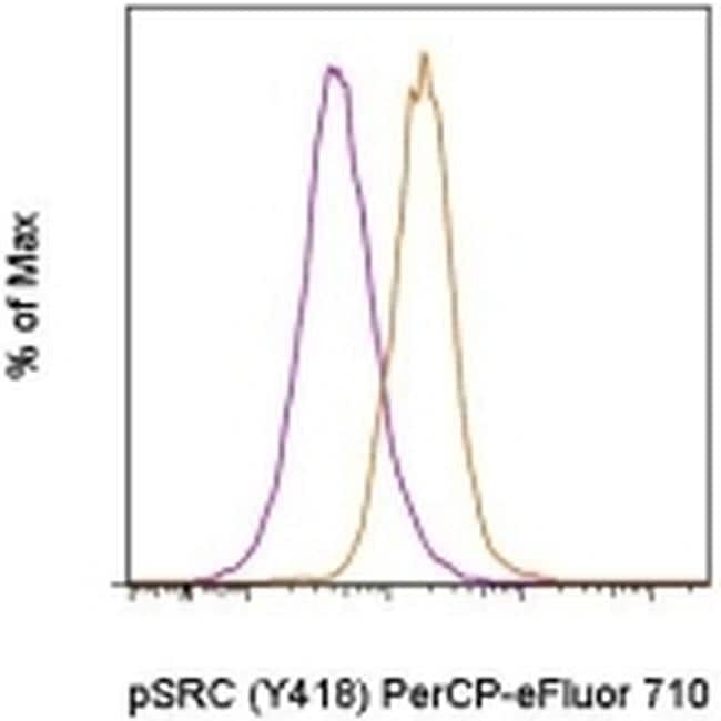
- Experimental details
- Intracellular staining of mouse spleen cells untreated (orange histogram) or treated with staurosporine for 2 hours (purple histogram) with Anti-Human/Mouse phospho-Src (Y418) PerCP-eFluor® 710 using the Intracellular Fixation & Permeabilization Buffer Set (Product # 88-8824-00) and protocol. Cells in the lymphocyte gate were used for analysis.
- Submitted by
- Invitrogen Antibodies (provider)
- Main image

- Experimental details
- Intracellular staining of mouse spleen cells untreated (orange histogram) or treated with staurosporine for 2 hours (purple histogram) with Anti-Human/Mouse phospho-Src (Y418) PerCP-eFluor® 710 using the Intracellular Fixation & Permeabilization Buffer Set (Product # 88-8824-00) and protocol. Cells in the lymphocyte gate were used for analysis.
