 Explore
 Validate
 Learn
Learn Western blot
 Immunocytochemistry
Antibody data
- Antibody Data
- Antigen structure
- References [0]
- Comments [0]
- Validations
- Western blot [1]
- Immunohistochemistry [5]
- Flow cytometry [1]
Submit
Validation data
Reference
Comment
Report error
- Product number
- NBP2-36429-0.1mg - Provider product page

- Provider
- Novus Biologicals
- Product name
- Mouse Monoclonal VEGFR2/KDR/Flk-1 Antibody
- Antibody type
- Monoclonal
- Description
- Protein G purified.
- Reactivity
- Human
- Host
- Mouse
- Isotype
- IgG
- Vial size
- 0.1 mg
- Concentration
- 1.0 mg/ml
- Storage
- Store at 4C short term. Aliquot and store at -20C long term. Avoid freeze-thaw cycles.
No comments: Submit comment
Supportive validation
- Submitted by
- Novus Biologicals (provider)
- Main image

- Experimental details
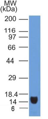
- Western Blot: VEGF R2/KDR/Flk-1 Antibody (2C6) [NBP2-36429] - Analysis of VEGF R2/KDR/Flk-1 (clone 2C6) in partial recombinant protein.
Supportive validation
- Submitted by
- Novus Biologicals (provider)
- Main image

- Experimental details
- Immunohistochemistry-Paraffin: VEGF R2/KDR/Flk-1 Antibody (2C6) [NBP2-36429] - Analysis of FFPE tissue section of Human SCC of Esophagus with antibody at 5 ug/ml concentration. The cancer cells depicted a specific and strong staining in the cytoplasm while the tumor stroma was negative for VEGF R2/KDR/Flk-1 immunopositivity.

- Submitted by
- Novus Biologicals (provider)
- Main image
- Experimental details
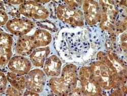
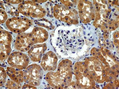
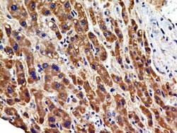
- Immunohistochemistry-Paraffin: VEGF R2/KDR/Flk-1 Antibody (2C6) [NBP2-36429] - Analysis of FFPE tissue section of human normal kidney with antibody at 5 ug/ml concentration. The cuboidal epithelial cells of of various tubules/ducts showed a specific cytoplasmic immunopositivity of VEGF R2/KDR/Flk-1 protein.
- Submitted by
- Novus Biologicals (provider)
- Main image

- Experimental details
- Immunohistochemistry-Paraffin: VEGF R2/KDR/Flk-1 Antibody (2C6) [NBP2-36429] - Analysis of FFPE tissue section of a malignant stromal tumor of the human small bowel with antibody at 5 ug/ml concentration. The cancer cells depicted a specific and strong staining in the cytoplasm.
- Submitted by
- Novus Biologicals (provider)
- Main image

- Experimental details
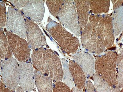
- Immunohistochemistry-Paraffin: VEGF R2/KDR/Flk-1 Antibody (2C6) [NBP2-36429] - Aanalysis of FFPE transverse section of normal human skeletal muscle with antibody at 5 ug/ml concentration. The myocytes showed a moderate diffused to granular staining in the cytoplasm.
- Submitted by
- Novus Biologicals (provider)
- Main image
- Experimental details
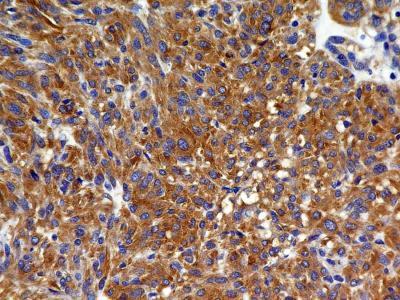
- Immunohistochemistry-Paraffin: VEGF R2/KDR/Flk-1 Antibody (2C6) [NBP2-36429] - Analysis of FFPE tissue section of Human Hepatocellular Carcinoma with antibody at 5 ug/ml concentration. The cancer cells depicted a specific and strong staining in the cytoplasm.

Supportive validation
- Submitted by
- Novus Biologicals (provider)
- Main image
- Experimental details
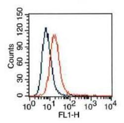
- Flow (Cell Surface): VEGF R2/KDR/Flk-1 Antibody (2C6) [NBP2-36429] - HepG2 cells were stained (surface) with antibody (orange) or isotype control, mouse IgG1 (blue). Positive staining observed using FITC conjugated mouse anti-IgG(H+L) secondary antibody. Live cells (PPI negative) were gated for analysis
