Explore
 Validate
 Learn
Learn Western blot
 ELISA
ELISAAntibody data
- Antibody Data
- Antigen structure
- References [0]
- Comments [0]
- Validations
- Western blot [2]
- Immunohistochemistry [1]
- Flow cytometry [2]
Submit
Validation data
Reference
Comment
Report error
- Product number
- NBP1-18646 - Provider product page

- Provider
- Novus Biologicals
- Proper citation
- Novus Cat#NBP1-18646, RRID:AB_1626056
- Product name
- Mouse Monoclonal VEGFR2/KDR/Flk-1 Antibody
- Antibody type
- Monoclonal
- Description
- Protein G purified. The monoclonal antibody will detect native human VEGFR-2/KDR in ELISA experiments and on the surface of different human cell types.
- Reactivity
- Human
- Host
- Mouse
- Isotype
- IgG
- Vial size
- 0.1 mg
- Concentration
- LYOPH
- Storage
- Store at 4C short term. Aliquot and store at -20C long term. Avoid freeze-thaw cycles.
No comments: Submit comment
Supportive validation
- Submitted by
- Novus Biologicals (provider)
- Main image
- Experimental details
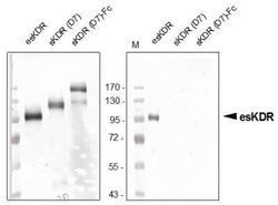
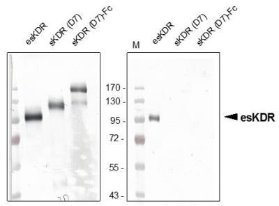
- Western Blot: VEGF R2/KDR/Flk-1 Antibody (20I6) [NBP1-18646] - Recombinant human endogenous soluble VEGFR-2/KDR (esKDR) was produced in insect cells. Western blot was performed using our monoclonal anti-VEGFR-2 recognizing the soluble as well as the transmembrane form of KDR (left panal) and our new poyclonal antibody directed against the unique C-terminal end of the endogenous sKDR (CGRETILDHSAEAVGMP) recognizing solely the endogenous form but not sKDR (D7) and sKDR (D7)-Fc consisting of the full extraplasmatic domain. The endogenous sKDR generated by alternative splicing consists of the first 6 Ig-like loops with a unique c-terminal end.
- Submitted by
- Novus Biologicals (provider)
- Main image
- Experimental details
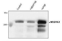
- Western Blot: VEGF R2/KDR/Flk-1 Antibody (20I6) [NBP1-18646] - Analysis of immunoprecipitated VEGFR-2/KDR form total lysate of HUVECs using anti-human VEGF Receptor 2

Supportive validation
- Submitted by
- Novus Biologicals (provider)
- Main image
- Experimental details
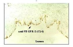
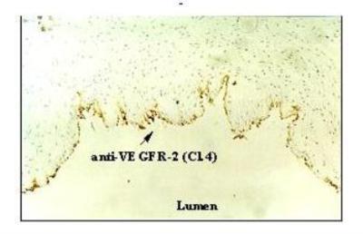
- Immunohistochemistry-Frozen: VEGF R2/KDR/Flk-1 Antibody (20I6) [NBP1-18646] - Fig. 4: Up-regulation of VEGFR-2/KDR in vein ECs of an intact human umbilical cord by FGF-2 (basic). A fresh human umbilical cord was rinsed with PBS to remove residual blood cells, cut in small pieces (about 0.5 cm), incubated in EBM (1% FCS) and stimulated with or without 20 ng/ml basic FGF for 24 h. Pieces were frozen in liquid nitrogen and used for immunohistochemistry using the mab anti-human VEGFR-2/Cl.4 as detection antibody.
Supportive validation
- Submitted by
- Novus Biologicals (provider)
- Main image
- Experimental details
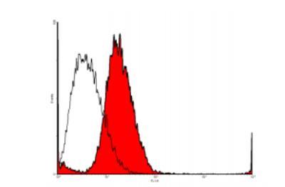
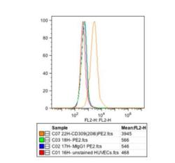
- Flow Cytometry: VEGF R2/KDR/Flk-1 Antibody (20I6) [NBP1-18646] - FACS analysis of VEGFR-2/KDR expression in HUVE cells [5ug/ml; 5ug/ml PE goat anti-mouse IgG]. The experiment was performed by Trisha M. Westerhof, University of California, Irvine.
- Submitted by
- Novus Biologicals (provider)
- Main image

- Experimental details
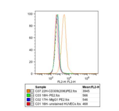
- Flow Cytometry: VEGF R2/KDR/Flk-1 Antibody (20I6) [NBP1-18646] - Analysis with primary human dermal lymphatic endothelial cells (HDLEC).