Explore
 Validate
 Learn
Learn Western blot
 Immunocytochemistry
Antibody data
- Antibody Data
- Antigen structure
- References [8]
- Comments [0]
- Validations
- Immunocytochemistry [4]
- Other assay [3]
Submit
Validation data
Reference
Comment
Report error
- Product number
- MA5-15157 - Provider product page

- Provider
- Invitrogen Antibodies
- Product name
- VEGF Receptor 2 Monoclonal Antibody (B.309.4)
- Antibody type
- Monoclonal
- Antigen
- Recombinant full-length protein
- Description
- It is not recommended to aliquot this antibody. This antibody is not cross-reactive with other family members.
- Reactivity
- Human, Mouse
- Host
- Rabbit
- Isotype
- IgG
- Antibody clone number
- B.309.4
- Vial size
- 100 μL
- Concentration
- 28 μg/mL
- Storage
- -20°C
Submitted references Cyclooxygenase inhibition attenuates brain angiogenesis and independently decreases mouse survival under hypoxia.
Functional damage of endothelial progenitor cells is attenuated by 14-3-3-n through inhibition of mitochondrial injury and oxidative stress.
VEGF Upregulates EGFR Expression to Stimulate Chemotactic Behaviors in the rMC-1 Model of Müller Glia.
Targeting QKI-7 in vivo restores endothelial cell function in diabetes.
Blockade of insulin receptor substrate-1 inhibits biological behavior of choroidal endothelial cells.
Inhibition of Endothelial SCUBE2 (Signal Peptide-CUB-EGF Domain-Containing Protein 2), a Novel VEGFR2 (Vascular Endothelial Growth Factor Receptor 2) Coreceptor, Suppresses Tumor Angiogenesis.
Generation of Hepatic Stellate Cells from Human Pluripotent Stem Cells Enables In Vitro Modeling of Liver Fibrosis.
Brain microvasculature defects and Glut1 deficiency syndrome averted by early repletion of the glucose transporter-1 protein.
Seeger DR, Golovko SA, Grove BD, Golovko MY
Journal of neurochemistry 2021 Jul;158(2):246-261
Journal of neurochemistry 2021 Jul;158(2):246-261
Functional damage of endothelial progenitor cells is attenuated by 14-3-3-n through inhibition of mitochondrial injury and oxidative stress.
Li Y, Sun X, Zhang X, Zhou H, Wang D, Xia Y, Li X
Cell biology international 2021 Apr;45(4):839-848
Cell biology international 2021 Apr;45(4):839-848
VEGF Upregulates EGFR Expression to Stimulate Chemotactic Behaviors in the rMC-1 Model of Müller Glia.
Peña JS, Vazquez M
Brain sciences 2020 May 29;10(6)
Brain sciences 2020 May 29;10(6)
Targeting QKI-7 in vivo restores endothelial cell function in diabetes.
Yang C, Eleftheriadou M, Kelaini S, Morrison T, González MV, Caines R, Edwards N, Yacoub A, Edgar K, Moez A, Ivetic A, Zampetaki A, Zeng L, Wilkinson FL, Lois N, Stitt AW, Grieve DJ, Margariti A
Nature communications 2020 Jul 30;11(1):3812
Nature communications 2020 Jul 30;11(1):3812
Blockade of insulin receptor substrate-1 inhibits biological behavior of choroidal endothelial cells.
Qian YY, Wu HY, Liu GQ, Ren C, Lu PR, Zhang XG
International journal of ophthalmology 2019;12(9):1386-1394
International journal of ophthalmology 2019;12(9):1386-1394
Inhibition of Endothelial SCUBE2 (Signal Peptide-CUB-EGF Domain-Containing Protein 2), a Novel VEGFR2 (Vascular Endothelial Growth Factor Receptor 2) Coreceptor, Suppresses Tumor Angiogenesis.
Lin YC, Liu CY, Kannagi R, Yang RB
Arteriosclerosis, thrombosis, and vascular biology 2018 May;38(5):1202-1215
Arteriosclerosis, thrombosis, and vascular biology 2018 May;38(5):1202-1215
Generation of Hepatic Stellate Cells from Human Pluripotent Stem Cells Enables In Vitro Modeling of Liver Fibrosis.
Coll M, Perea L, Boon R, Leite SB, Vallverdú J, Mannaerts I, Smout A, El Taghdouini A, Blaya D, Rodrigo-Torres D, Graupera I, Aguilar-Bravo B, Chesne C, Najimi M, Sokal E, Lozano JJ, van Grunsven LA, Verfaillie CM, Sancho-Bru P
Cell stem cell 2018 Jul 5;23(1):101-113.e7
Cell stem cell 2018 Jul 5;23(1):101-113.e7
Brain microvasculature defects and Glut1 deficiency syndrome averted by early repletion of the glucose transporter-1 protein.
Tang M, Gao G, Rueda CB, Yu H, Thibodeaux DN, Awano T, Engelstad KM, Sanchez-Quintero MJ, Yang H, Li F, Li H, Su Q, Shetler KE, Jones L, Seo R, McConathy J, Hillman EM, Noebels JL, De Vivo DC, Monani UR
Nature communications 2017 Jan 20;8:14152
Nature communications 2017 Jan 20;8:14152
No comments: Submit comment
Supportive validation
- Submitted by
- Invitrogen Antibodies (provider)
- Main image
- Experimental details
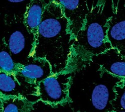
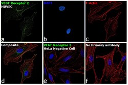
- Immunofluorescent analysis of VEGF Receptor 2 in HUVEC cells using a VEGF Receptor 2 monoclonal antibody (Product # MA5-15157) (green). DNA is labeled using a fluorescent blue dye.
- Submitted by
- Invitrogen Antibodies (provider)
- Main image
- Experimental details
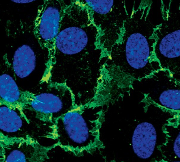
- Immunofluorescent analysis of VEGF Receptor 2 in HUVEC cells, stimulated with Vascular Endothelial Growth Factor, using a VEGF Receptor 2 monoclonal antibody (Product # MA5-15157) (green). DNA is labeled using a fluorescent blue dye.

- Submitted by
- Invitrogen Antibodies (provider)
- Main image
- Experimental details
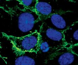
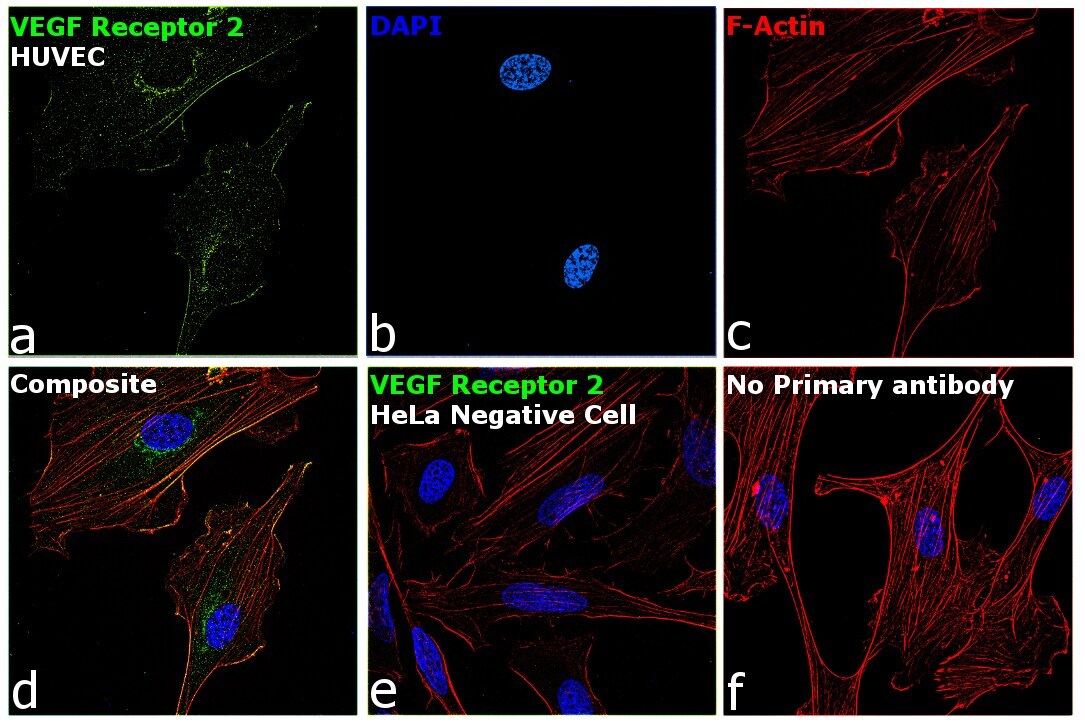
- Immunofluorescence analysis of VEGF Receptor 2 was performed using 70% confluent log phase HUVEC cells. The cells were fixed with 4% paraformaldehyde for 10 minutes, permeabilized with 0.1% Triton™ X-100 for 15 minutes, and blocked with 2% BSA for 45 minutes at room temperature. The cells were labeled with VEGF Receptor 2 Monoclonal Antibody (B.309.4) (Product # MA5-15157) at 1:200 dilution in 0.1% BSA, incubated at 4 degree celsius overnight and then labeled with Donkey anti-Mouse IgG (H+L) Highly Cross-Adsorbed Secondary Antibody, Alexa Fluor Plus 488 (Product # A32766), (1:2000 dilution), for 45 minutes at room temperature (Panel a: Green). Nuclei (Panel b: Blue) were stained with ProLong™ Diamond Antifade Mountant with DAPI (Product # P36962). F-actin (Panel c: Red) was stained with Rhodamine Phalloidin (Product # R415, 1:300). Panel d represents the merged image showing Plasma Membrane and Endoplasmic Reticulum localization. Panel e represents HeLa cells with no expression of VEGF Receptor 2. Panel f represents control cells with no primary antibody to assess background. The images were captured at 60X magnification.
- Submitted by
- Invitrogen Antibodies (provider)
- Main image

- Experimental details
- Immunofluorescence analysis of VEGF Receptor 2 was performed using 70% confluent log phase HUVEC cells. The cells were fixed with 4% paraformaldehyde for 10 minutes, permeabilized with 0.1% Triton™ X-100 for 15 minutes, and blocked with 2% BSA for 45 minutes at room temperature. The cells were labeled with VEGF Receptor 2 Monoclonal Antibody (B.309.4) (Product # MA5-15157) at 1:200 dilution in 0.1% BSA, incubated at 4 degree celsius overnight and then labeled with Donkey anti-Mouse IgG (H+L) Highly Cross-Adsorbed Secondary Antibody, Alexa Fluor Plus 488 (Product # A32766), (1:2000 dilution), for 45 minutes at room temperature (Panel a: Green). Nuclei (Panel b: Blue) were stained with ProLong™ Diamond Antifade Mountant with DAPI (Product # P36962). F-actin (Panel c: Red) was stained with Rhodamine Phalloidin (Product # R415, 1:300). Panel d represents the merged image showing Plasma Membrane and Endoplasmic Reticulum localization. Panel e represents HeLa cells with no expression of VEGF Receptor 2. Panel f represents control cells with no primary antibody to assess background. The images were captured at 60X magnification.

Supportive validation
- Submitted by
- Invitrogen Antibodies (provider)
- Main image

- Experimental details
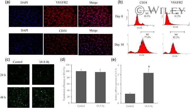
- 1 Figure Endothelial progenitor cells (EPCs) were identified using CD34 and VEGFR2, and overexpression of 14-3-3-eta in the EPCs were established. (a) Identification of EPCs by detecting VEGFR2 and CD34. (b) Identification of VEGFR2 and CD34 in EPCs through flow cytometry. (c) Transfection of 14-3-3-eta overexpression vector into EPCs. (d) Transfection efficiency of 4-3-3-eta overexpression vector was measured. (e) Relative mRNA expression of 14-3-3-eta in the EPCs was measured after transfection. * p < .05 compared with control group. Experiments were repeated independently three times
- Submitted by
- Invitrogen Antibodies (provider)
- Main image
- Experimental details
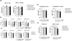
- FIGURE 5 Alterations in brain angiogenic factor under hypoxia and COX inhibition with ketorolac. Mice were treated with saline (1 and 2) or ketorolac (3 and 4) using osmotic pumps (s.c.) under 24-hr or 10-day normoxia (1 and 3, 20% O 2 ) or hypoxia (2 and 4, 10% O 2 ). Values are mean +- standard deviation ( n (number of animals) =4) with individual values. Values that do not share the same letter are statistically different ( p < .05, one-way ANOVA with Tukey's post hoc test). (a) COX inhibition with ketorolac has no effect on HIF-1alpha levels under brain hypoxia as determined using ELISA. (b) COX inhibition with ketorolac has a slight but significant effect on brain VEGF and VEGFR2 levels under hypoxia. Brain VEGF was determined using ELISA. VEGFR2 were determined after western blot analysis and relative optical densities against actin are presented. Panels below bar graphs show representative blots for VEGFR2. (c). COX inhibition with ketorolac has no effect on brain bFGF and FGFR1 levels under hypoxia. Brain bFGF was determined using ELISA. pFGFR1 was determined after western blot analysis and relative optical densities against actin are presented. Panels below bar graphs show representative blots for pFGFR1. (d) COX inhibition with ketorolac has no effect on brain angiopoietin-2 under hypoxia. Brain angiopoietin-2 (Ang-2) was determined after western blot analysis and expressed as relative optical densities against actin. Panels below bar graphs show representative blot

- Submitted by
- Invitrogen Antibodies (provider)
- Main image

- Experimental details
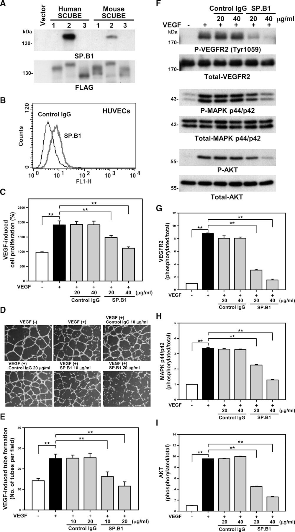
- Figure 3. Anti-SCUBE2 (signal peptide-CUB-EGF domain-containing protein 2) monoclonal antibody (mAb) SP.B1 inhibits VEGF (vascular endothelial growth factor)-induced responses and signal transduction in human umbilical vein ECs (HUVECs). A , B , Characterization of anti-SCUBE2 mAb SP.B1. Western blot analysis and flow cytometry of mAb SP.B1 recognizing human and mouse recombinant SCUBE2 but not 1 and 3 protein expressed in HEK-293T cells ( A ) and cell surface expression of SCUBE2 in HUVECs ( B ). C - I , The mAb SP.B1 blocks VEGF-stimulated cell responses and VEGF-induced VEGFR2 (VEGF receptor 2) phosphorylation and MAPK (mitogen-activated protein kinase)&solAKT activation. VEGF-induced HUVEC proliferation ( C ) and angiogenesis ( D , E ) with the addition of SP.B1 and control IgG. Western blot analysis of VEGF-induced phosphorylated and total VEGFR2, MAPK, and AKT in HUVECs with SP.B1 or control IgG treatment ( F ) and quantification ( G - I ). Data are mean+-SD from 3 independent experiments ( G - I ). &ast&ast P