Explore
 Validate
 Learn
Learn Western blot
 Immunohistochemistry
Antibody data
- Antibody Data
- Antigen structure
- References [8]
- Comments [0]
- Validations
- Immunohistochemistry [1]
- Other assay [5]
Submit
Validation data
Reference
Comment
Report error
- Product number
- PA5-16487 - Provider product page

- Provider
- Invitrogen Antibodies
- Product name
- VEGF Receptor 2 Polyclonal Antibody
- Antibody type
- Polyclonal
- Antigen
- Synthetic peptide
- Description
- PA5-16487 targets Flk-1 in IHC (P) and WB applications and shows reactivity with mouse, Rat, and Human samples. The PA5-16487 immunogen is a synthetic peptide from C-terminus of the precursor form of the mouse Flk-1.
- Reactivity
- Human, Mouse, Rat
- Host
- Rabbit
- Isotype
- IgG
- Vial size
- 500 μL
- Concentration
- 1 mg/mL
- Storage
- 4°C
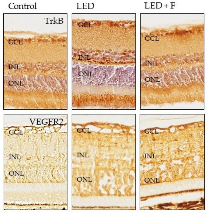
Submitted references Retinal Protection from LED-Backlit Screen Lights by Short Wavelength Absorption Filters.
In vitro angiogenesis inhibition with selective compounds targeting the key glycolytic enzyme PFKFB3.
SARS-CoV-2 spike protein co-opts VEGF-A/neuropilin-1 receptor signaling to induce analgesia.
SARS-CoV-2 Spike protein co-opts VEGF-A/Neuropilin-1 receptor signaling to induce analgesia.
Hypoxia and proangiogenic proteins in human ameloblastoma.
A phase II study of tivozanib in patients with metastatic and nonresectable soft-tissue sarcomas.
Myoferlin expression in non-small cell lung cancer: Prognostic role and correlation with VEGFR-2 expression.
Expression profiling and significance of VEGF-A, VEGFR2, VEGFR3 and related proteins in endometrial carcinoma.
Sanchez-Ramos C, Bonnin-Arias C, Blázquez-Sánchez V, Aguirre-Vilacoro V, Cobo T, García-Suarez O, Perez-Carrasco MJ, Alvarez-Peregrina C, Vega JA
Cells 2021 Nov 19;10(11)
Cells 2021 Nov 19;10(11)
In vitro angiogenesis inhibition with selective compounds targeting the key glycolytic enzyme PFKFB3.
Abdali A, Baci D, Damiani I, Belloni F, De Dominicis C, Gelmi ML, Corsini A, Bellosta S
Pharmacological research 2021 Jun;168:105592
Pharmacological research 2021 Jun;168:105592
SARS-CoV-2 spike protein co-opts VEGF-A/neuropilin-1 receptor signaling to induce analgesia.
Moutal A, Martin LF, Boinon L, Gomez K, Ran D, Zhou Y, Stratton HJ, Cai S, Luo S, Gonzalez KB, Perez-Miller S, Patwardhan A, Ibrahim MM, Khanna R
Pain 2021 Jan;162(1):243-252
Pain 2021 Jan;162(1):243-252
SARS-CoV-2 Spike protein co-opts VEGF-A/Neuropilin-1 receptor signaling to induce analgesia.
Moutal A, Martin LF, Boinon L, Gomez K, Ran D, Zhou Y, Stratton HJ, Cai S, Luo S, Gonzalez KB, Perez-Miller S, Patwardhan A, Ibrahim MM, Khanna R
bioRxiv : the preprint server for biology 2020 Sep 14;
bioRxiv : the preprint server for biology 2020 Sep 14;
Hypoxia and proangiogenic proteins in human ameloblastoma.
de Mendonça RP, Balbinot KM, Martins BV, da Silva Kataoka MS, Mesquita RA, de Jesus Viana Pinheiro J, de Melo Alves Júnior S
Scientific reports 2020 Oct 16;10(1):17567
Scientific reports 2020 Oct 16;10(1):17567
A phase II study of tivozanib in patients with metastatic and nonresectable soft-tissue sarcomas.
Agulnik M, Costa RLB, Milhem M, Rademaker AW, Prunder BC, Daniels D, Rhodes BT, Humphreys C, Abbinanti S, Nye L, Cehic R, Polish A, Vintilescu C, McFarland T, Skubitz K, Robinson S, Okuno S, Van Tine BA
Annals of oncology : official journal of the European Society for Medical Oncology 2017 Jan 1;28(1):121-127
Annals of oncology : official journal of the European Society for Medical Oncology 2017 Jan 1;28(1):121-127
Myoferlin expression in non-small cell lung cancer: Prognostic role and correlation with VEGFR-2 expression.
Song DH, Ko GH, Lee JH, Lee JS, Lee GW, Kim HC, Yang JW, Heo RW, Roh GS, Han SY, Kim DC
Oncology letters 2016 Feb;11(2):998-1006
Oncology letters 2016 Feb;11(2):998-1006
Expression profiling and significance of VEGF-A, VEGFR2, VEGFR3 and related proteins in endometrial carcinoma.
Wang J, Taylor A, Showeil R, Trivedi P, Horimoto Y, Bagwan I, Ewington L, Lam EW, El-Bahrawy MA
Cytokine 2014 Aug;68(2):94-100
Cytokine 2014 Aug;68(2):94-100
No comments: Submit comment
Supportive validation
- Submitted by
- Invitrogen Antibodies (provider)
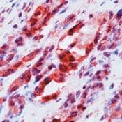
- Main image

- Experimental details
- Formalin-fixed, paraffin-embedded human angiosarcoma stained with Flk-1/ VEGFR2 antibody using peroxidase-conjugate and AEC chromogen. Note cytoplasmic staining of tumor cells.
Supportive validation
- Submitted by
- Invitrogen Antibodies (provider)
- Main image

- Experimental details
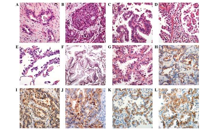
- Figure 2. Microscopic features of adenocarcinoma following hematoxylin and eosin staining. (A) Acinar pattern of adenocarcinoma revealed glands with central lumina. (B) Solid pattern adenocarcinoma revealed highly packed cells with no specific pattern. (C) Papillary pattern adenocarcinoma exhibited tumor cell proliferation with a central vascular core. (D) Micropapillary pattern adenocarcinoma revealed projective cell proliferation with no fibrovascular core. (E) Lepidic pattern adenocarcinoma revealed a preserved alveolar structure. (F) Proliferation of mucin-containing tumor cells was observed in mucinous adenocarcinoma. (G) Adenocarcinoma without thyroid transcription factor-1 staining and nuclear pleomorphism with occasionally prominent nucleoli. This tumor was classified as low nuclear grade. (H) Cytoplasmic myoferlin expression. (I) Vascular endothelial growth factor receptor-2 was additionally expressed in the cytoplasm. (J) E-cadherin protein was located in the cell membrane with focal expression in the cytoplasm. (K) beta-catenin was expressed in the cell membrane. (L) Epidermal growth factor receptor protein was additionally expressed in the cell membrane. Magnification, x400.
- Submitted by
- Invitrogen Antibodies (provider)
- Main image

- Experimental details
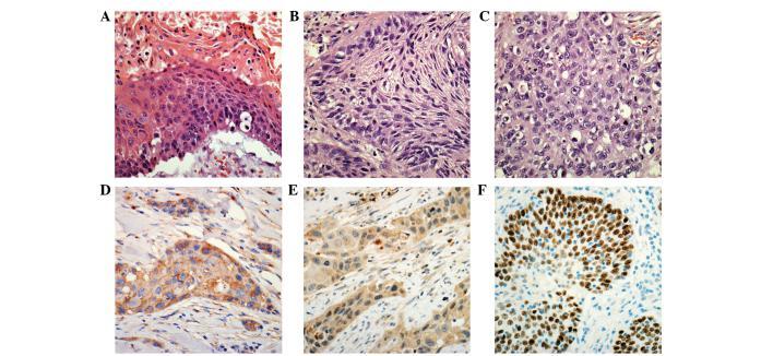
- Figure 1. Microscopic features of squamous cell carcinoma following hematoxylin and eosin staining. (A) Well-differentiated squamous cell carcinoma demonstrated keratinization with parakeratosis and a relatively distinct cell border. (B) Moderately-differentiated squamous cell carcinoma demonstrated a distinct cell border, however, no keratinization was observed. (C) Poorly-differentiated squamous cell carcinoma exhibited a vague cellular border. This tumor was positive for p63 immunostaining and negative for thyroid transcription factor-1 immunostaining, indicating a differential diagnosis from solid pattern adenocarcinoma. (D) Myoferlin protein was expressed in the cytoplasm of the tumor cells. Capillary endothelial cells in the stroma additionally demonstrated myoferlin expression. (E) Vascular endothelial growth factor receptor-2 expression was observed in the cytoplasm of the tumor cells. (F) Poorly-differentiated squamous cell carcinoma demonstrated p63 expression in the nucleus. Magnification, x400.
- Submitted by
- Invitrogen Antibodies (provider)
- Main image

- Experimental details
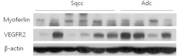
- Figure 3. Western blot analysis of non-small cell carcinoma samples from 6 patients with squamous cell carcinoma and four patients with adenocarcinoma. Immunohistochemically, all patients demonstrated positive expression for myoferlin protein. Upon western blotting, there were also positive signals for myoferlin. Several specimens additionally demonstrated VEGFR-2 expression. beta-actin served as a control for equivalent protein loading. Sqcc, squamous cell carcinoma; Adc, adenocarcinoma; VEGFR-2, vascular endothelial growth factor receptor-2.
- Submitted by
- Invitrogen Antibodies (provider)
- Main image

- Experimental details
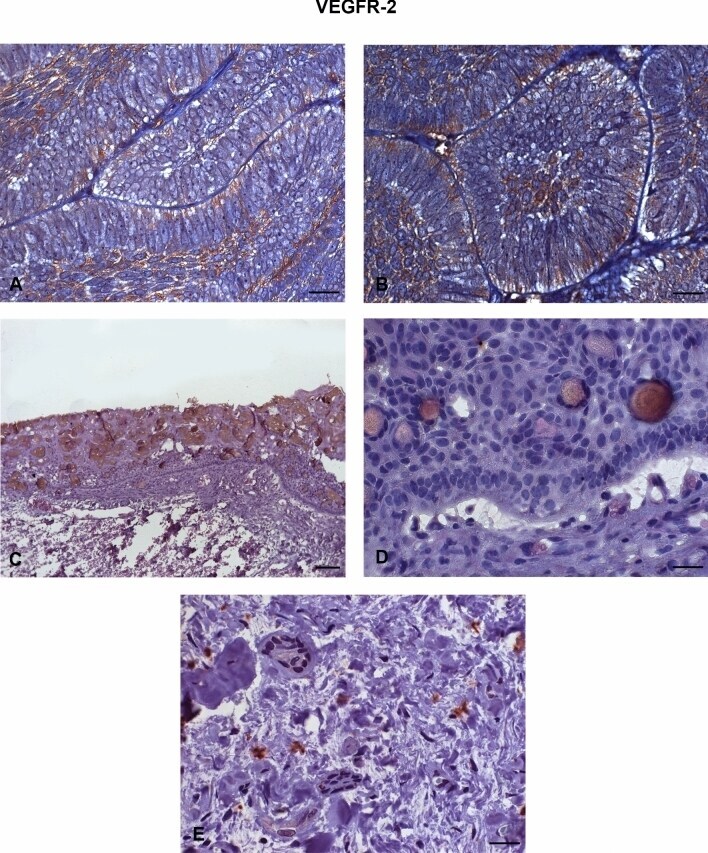
- Figure 4 Immunostaining of the vascular endothelial growth factor receptor (VEGFR-2) in ameloblastomas (AMEs), calcifying odontogenic cysts (COCs), and dental follicles (DFs). ( A , B ) Strong immunostaining in the cell membrane of ameloblastoma tumour cells. ( C , D ) Low intensity immunoexpression in a calcifying odontogenic cyst. ( E ) Low immunoexpression in a dental follicle. Immunoperoxidase. Scale bar: ( A , B , D , E ) 20 um; ( c ) 100 um.
- Submitted by
- Invitrogen Antibodies (provider)
- Main image

- Experimental details
- Figure 9 Immunohistochemical detection of TrkB ( upper panel ), and VEGFR2 ( lower panel ) in the three groups of rats investigated. LED: animals exposed to LED-backlit screen for 3 months; LED + F: animals exposed to filtered LED-backlit screen for 3 months. GCL: ganglionic cells layer; INL: inner nuclear layer; ONL: outer nuclear layer.