Explore
 Validate
 Learn
Learn Western blot
 ELISA
ELISA Immunocytochemistry
Antibody data
- Antibody Data
- Antigen structure
- References [3]
- Comments [0]
- Validations
- Immunocytochemistry [2]
- Flow cytometry [2]
- Other assay [1]
Submit
Validation data
Reference
Comment
Report error
- Product number
- MA5-15556 - Provider product page

- Provider
- Invitrogen Antibodies
- Product name
- VEGF Receptor 2 Monoclonal Antibody (4B4)
- Antibody type
- Monoclonal
- Antigen
- Purifed from natural sources
- Description
- MA5-15556 targets KDR in indirect ELISA, FACS, IF, and WB applications and shows reactivity with Human samples. The MA5-15556 immunogen is purified recombinant extracellular fragment of human KDR (aa20-764) fused with hIgGFc tag expressed in HEK293 cells. MA5-15556 detects KDR which has a predicted molecular weight of approximately 152kDa.
- Reactivity
- Human
- Host
- Mouse
- Isotype
- IgG
- Antibody clone number
- 4B4
- Vial size
- 100 μL
- Concentration
- Conc. Not Determined
- Storage
- Store at 4°C short term. For long term storage, store at -20°C, avoiding freeze/thaw cycles.
Submitted references Myocyte Enhancer Factor 2C as a New Player in Human Breast Cancer Brain Metastases.
Carcinoma cells that have undergone an epithelial-mesenchymal transition differentiate into endothelial cells and contribute to tumor growth.
A new algorithm for a better characterization and timing of the anti-VEGF vascular effect named "normalization".
Galego S, Kauppila LA, Malhó R, Pimentel J, Brito MA
Cells 2021 Feb 12;10(2)
Cells 2021 Feb 12;10(2)
Carcinoma cells that have undergone an epithelial-mesenchymal transition differentiate into endothelial cells and contribute to tumor growth.
Sphyris N, King C, Hoar J, Werden SJ, Vijay GV, Miura N, Gaharwar A, Sarkar TR
Oncotarget 2021 Apr 13;12(8):823-844
Oncotarget 2021 Apr 13;12(8):823-844
A new algorithm for a better characterization and timing of the anti-VEGF vascular effect named "normalization".
El Alaoui-Lasmaili K, Djermoune EH, Tylcz JB, Meng D, Plénat F, Thomas N, Faivre B
Angiogenesis 2017 Feb;20(1):149-162
Angiogenesis 2017 Feb;20(1):149-162
No comments: Submit comment
Supportive validation
- Submitted by
- Invitrogen Antibodies (provider)
- Main image

- Experimental details
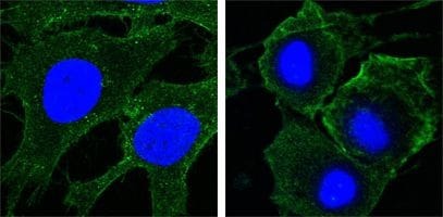
- Immunofluorescence analysis of HeLa (left) and HepG2 (right) cells using CD309/VEGFR2 monoclonal antibody (Product # MA5-15556) (Green). Blue: DRAQ5 fluorescent DNA dye.
- Submitted by
- Invitrogen Antibodies (provider)
- Main image

- Experimental details
- Immunofluorescence analysis of HeLa (left) and HepG2 (right) cells using CD309/VEGFR2 monoclonal antibody (Product # MA5-15556) (Green). Blue: DRAQ5 fluorescent DNA dye.

Supportive validation
- Submitted by
- Invitrogen Antibodies (provider)
- Main image

- Experimental details
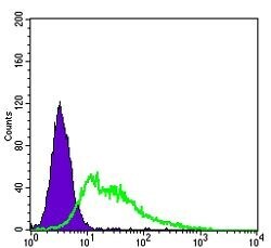
- Flow cytometric analysis of HepG2 cells using CD309/VEGFR2 monoclonal antibody (Product # MA5-15556) (green) and negative control (purple).
- Submitted by
- Invitrogen Antibodies (provider)
- Main image

- Experimental details
- Flow cytometric analysis of HepG2 cells using CD309/VEGFR2 monoclonal antibody (Product # MA5-15556) (green) and negative control (purple).

Supportive validation
- Submitted by
- Invitrogen Antibodies (provider)
- Main image

- Experimental details
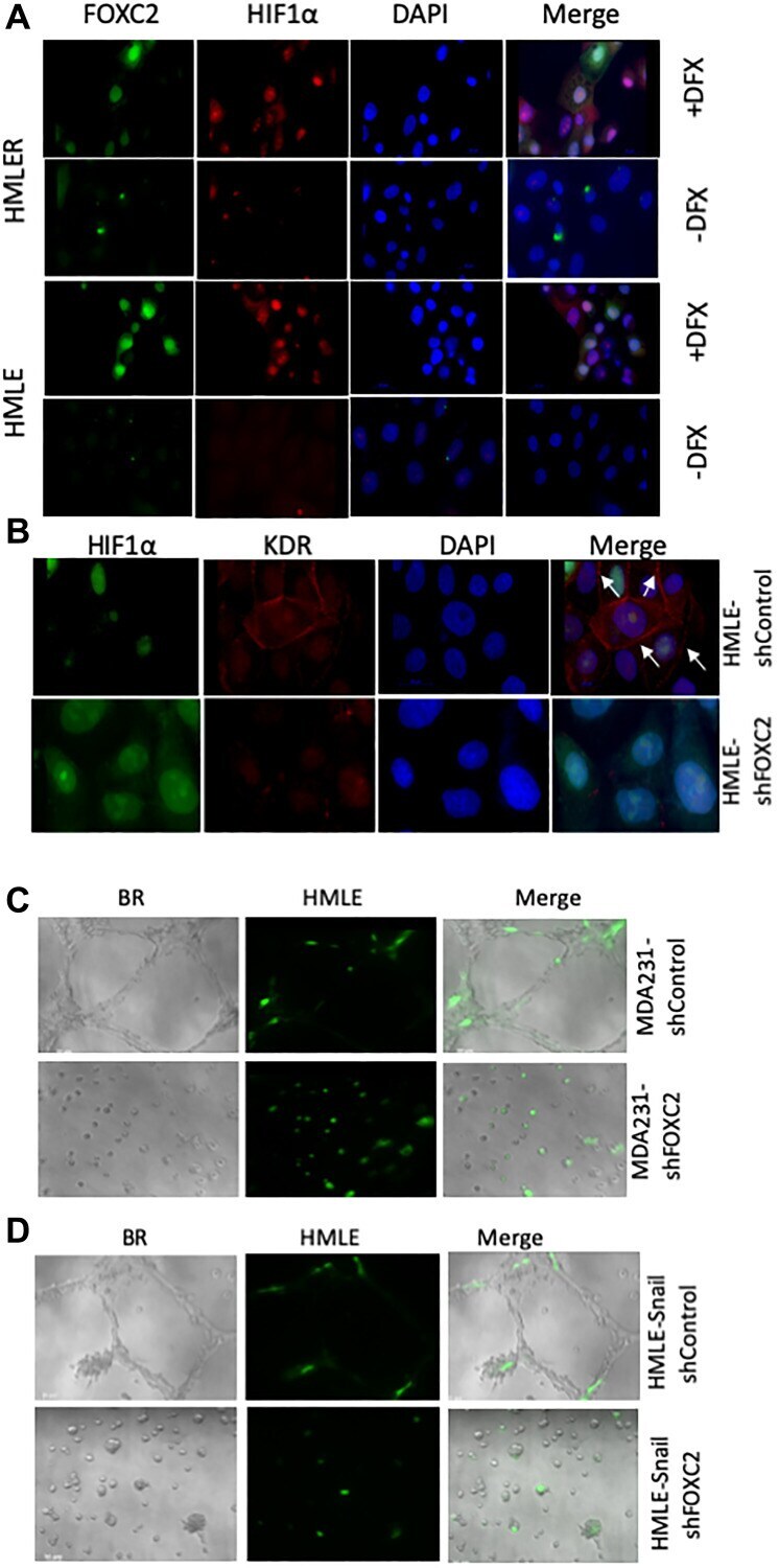
- Figure 6 FOXC2 is necessary for the acquisition of endothelial phenotypic and functional characteristics in vitro . ( A ) Immortalized (HMLE) and RAS-transformed (HMLER) human mammary epithelial cells were plated in cell-specific medium. After 24 hours, the cells were treated either with vehicle or desferrioxamine (DFX), a hypoxia-mimetic, for 48 hours prior to fixation and immunostaining for FOXC2 (green) and HIF-1alpha (red). Nuclei were counterstained with DAPI (blue). Right panels are merged images of individual channels. Scale bar: 20 mum. ( B ) HMLE cells, transduced with shRNAs targeting firefly luciferase (shControl) or FOXC2 (shFOXC2), were treated with DFX for 48 hours. Following DFX treatment, the cells were fixed and immunostained for HIF-1alpha (green) and KDR (red). Nuclei were counterstained with DAPI (blue). Right panels are merged images of individual channels. Scale bar: 20 mum ( C ) GFP-labeled HMLE cells (green) were co-mixed with either MDA-MB-231-shControl or MDA-MB-231-shFOXC2 cells and plated onto Matrigel in EGM-2. After 24 hours, the cells were imaged using brightfield (BR) and fluorescence microscopy. Right panels are overlays of brightfield and fluorescent images. The impact of FOXC2 knockdown on the ability of MDA-MB-231 cells to form vascular-like networks and stimulate the incorporation of GFP-labeled HMLE cells into mosaic structures was examined. ( D ) GFP-labeled HMLE cells (green) were co-mixed with either HMLE-Snail-shControl or HMLE-Snail-