 Explore
 Validate
 Learn
Learn Western blot
Antibody data
- Antibody Data
- Antigen structure
- References [5]
- Comments [0]
- Validations
- Western blot [1]
- Immunocytochemistry [1]
- Flow cytometry [1]
Submit
Validation data
Reference
Comment
Report error
- Product number
- AF1766 - Provider product page

- Provider
- R&D Systems
- Product name
- Human Phospho-VEGFR2/KDR/Flk-1 (Y1214) Antibody
- Antibody type
- Polyclonal
- Description
- Antigen Affinity-purified. Detects human VEGFR2/KDR/Flk-1 in Western blots when phosphorylated at Y1214.
- Reactivity
- Human
- Host
- Rabbit
- Conjugate
- Unconjugated
- Isotype
- IgG
- Vial size
- 50 ug
- Concentration
- LYOPH
- Storage
- Use a manual defrost freezer and avoid repeated freeze-thaw cycles. 12 months from date of receipt, -20 to -70 °C as supplied. 1 month, 2 to 8 °C under sterile conditions after reconstitution. 6 months, -20 to -70 °C under sterile conditions after reconstitution.
Submitted references Upregulation of RASSF1A in Colon Cancer by Suppression of Angiogenesis Signaling and Akt Activation.
Endothelial progenitor cells transplantation attenuated blood-brain barrier damage after ischemia in diabetic mice via HIF-1α.
DSGOST inhibits tumor growth by blocking VEGF/VEGFR2-activated angiogenesis.
Fluorescent resonance energy transfer imaging of VEGFR dimerization.
Vascular endothelial growth factor signals through platelet-derived growth factor receptor β in meningiomas in vitro.
Blanchard TG, Lapidus R, Banerjee V, Bafford AC, Czinn SJ, Ahmed H, Banerjee A
Cellular physiology and biochemistry : international journal of experimental cellular physiology, biochemistry, and pharmacology 2018;48(3):1259-1273
Cellular physiology and biochemistry : international journal of experimental cellular physiology, biochemistry, and pharmacology 2018;48(3):1259-1273
Endothelial progenitor cells transplantation attenuated blood-brain barrier damage after ischemia in diabetic mice via HIF-1α.
Geng J, Wang L, Qu M, Song Y, Lin X, Chen Y, Mamtilahun M, Chen S, Zhang Z, Wang Y, Yang GY
Stem cell research & therapy 2017 Jul 11;8(1):163
Stem cell research & therapy 2017 Jul 11;8(1):163
DSGOST inhibits tumor growth by blocking VEGF/VEGFR2-activated angiogenesis.
Choi HS, Lee K, Kim MK, Lee KM, Shin YC, Cho SG, Ko SG
Oncotarget 2016 Apr 19;7(16):21775-85
Oncotarget 2016 Apr 19;7(16):21775-85
Fluorescent resonance energy transfer imaging of VEGFR dimerization.
Ahmadova Z, Yagublu V, Förg T, Hajiyeva Y, Jesenofsky R, Hafner M, Keese M
Anticancer research 2014 May;34(5):2123-33
Anticancer research 2014 May;34(5):2123-33
Vascular endothelial growth factor signals through platelet-derived growth factor receptor β in meningiomas in vitro.
Pfister C, Pfrommer H, Tatagiba MS, Roser F
British journal of cancer 2012 Nov 6;107(10):1702-13
British journal of cancer 2012 Nov 6;107(10):1702-13
No comments: Submit comment
Supportive validation
- Submitted by
- R&D Systems (provider)
- Main image
- Experimental details
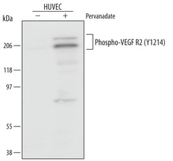
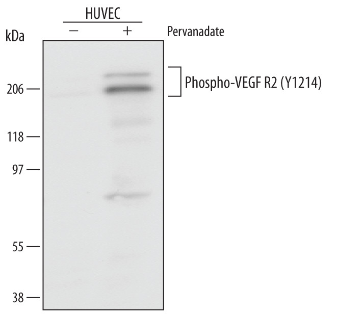
- Detection of Human Phospho-VEGFR2/KDR/Flk-1 (Y1214) by Western Blot. Western blot shows lysates of HUVEC human umbilical vein endothelial cells untreated (-) or treated (+) with 100 μM pervanadate (PV) for 10 minutes. PVDF membrane was probed with 0.5 µg/mL of Rabbit Anti-Human Phospho-VEGFR2/KDR/Flk-1 (Y1214) Antigen Affinity-purified Polyclonal Antibody, followed by HRP-conjugated Anti-Rabbit IgG Secondary Antibody (Catalog # HAF008). Specific bands were detected for Phospho-VEGFR2/KDR/Flk-1 (Y1214) at approximately 210 kDa (as indicated). This experiment was conducted under reducing conditions and using Immunoblot Buffer Group 1.
Supportive validation
- Submitted by
- R&D Systems (provider)
- Main image
- Experimental details
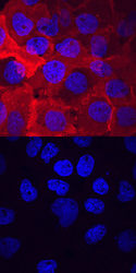
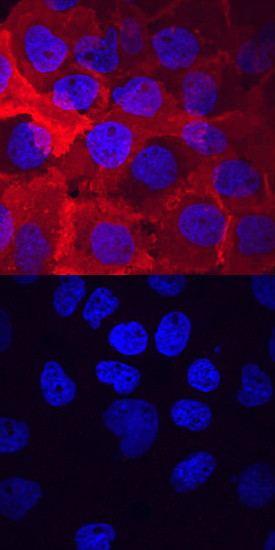
- Phospho-VEGFR2/KDR/Flk-1 (Y1214) in A431 Human Cell Line. VEGFR2/KDR/Flk-1 phosphorylated at Y1214 was detected in immersion fixed A431 human epithelial carcinoma cell line untreated (lower panel) or treated (upper panel) with pervanadate using Rabbit Anti-Human Phospho-VEGFR2/KDR/Flk-1 (Y1214) Antigen Affinity-purified Polyclonal Antibody (Catalog # AF1766) at 10 µg/mL for 3 hours at room temperature. Cells were stained using the NorthernLights™ 557-conjugated Anti-Rabbit IgG Secondary Antibody (red; Catalog # NL004) and counterstained with DAPI (blue). View our protocol for Fluorescent ICC Staining of Cells on Coverslips.
Supportive validation
- Submitted by
- R&D Systems (provider)
- Main image
- Experimental details
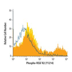
- Detection of Phospho-VEGFR2/KDR/Flk-1 in pervanadate-treated HUVEC by Flow Cytometry. HUVEC human umbilical vein endothelial cells were unstimulated (yellow filled histogram) or treated with 100 μM pervanadate for 15 minutes (orange filled histogram), then stained with Rabbit Anti-Human Phospho-VEGFR2/KDR/Flk-1 (Y1214) Antigen Affinity-purified Polyclonal Antibody (Catalog # AF1766), or control antibody (Catalog # AB-105-C, open histogram), followed by Phycoerythrin-conjugated Anti-Rabbit IgG Secondary Antibody (Catalog # F0110). To facilitate intracellular staining, cells were fixed with paraformaldehyde and permeabilized with methanol.
