Explore
 Validate
 Learn
Learn Western blot
Antibody data
- Antibody Data
- Antigen structure
- References [0]
- Comments [0]
- Validations
- Western blot [8]
- Immunocytochemistry [2]
Submit
Validation data
Reference
Comment
Report error
- Product number
- MA5-24966 - Provider product page

- Provider
- Invitrogen Antibodies
- Product name
- IDH1 Monoclonal Antibody (OTI2H9)
- Antibody type
- Monoclonal
- Antigen
- Other
- Reactivity
- Human, Mouse
- Host
- Mouse
- Isotype
- IgG
- Antibody clone number
- OTI2H9
- Vial size
- 100 μL
- Concentration
- 1 mg/mL
- Storage
- -20°C, Avoid Freeze/Thaw Cycles
No comments: Submit comment
Supportive validation
- Submitted by
- Invitrogen Antibodies (provider)
- Main image
- Experimental details
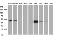
- Western blot analysis of IDH1 in human tissue (1: Testis; 2: Omentum; 3: Uterus; 4: Breast; 5: Brain; 6: Liver; 7: Ovary; 8: Thyroid gland; 9: Colon) samples using 10 µg per lane. Samples were separated by SDS-PAGE and probed with IDH1 (Product # MA5-24966) monoclonal antibody at a dilution of 1:200.
- Submitted by
- Invitrogen Antibodies (provider)
- Main image
- Experimental details
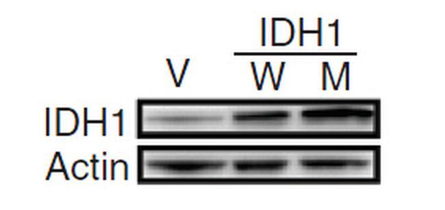
- Western blot analysis of IDH1 in HEK293T cells in untransfected (Left lane) and transfected (Right lane) samples using 5 µg per lane. The samples were separated by SDS-PAGE and probed with IDH1 (Product # MA5-24966) monoclonal antibody.
- Submitted by
- Invitrogen Antibodies (provider)
- Main image
- Experimental details
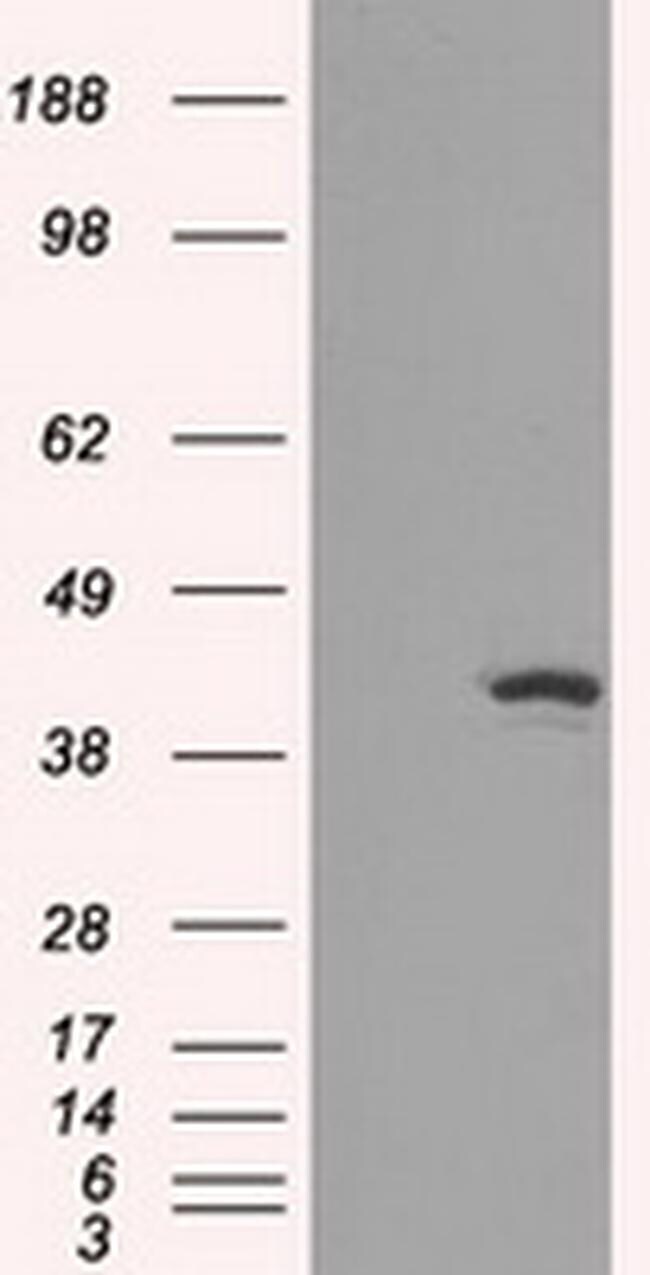
- Western blot analysis of IDH1 in normal human astrocyte tissue. Samples were separated by SDS-PAGE and probed with IDH1 (Product # MA5-24966) monoclonal antibody.
- Submitted by
- Invitrogen Antibodies (provider)
- Main image

- Experimental details
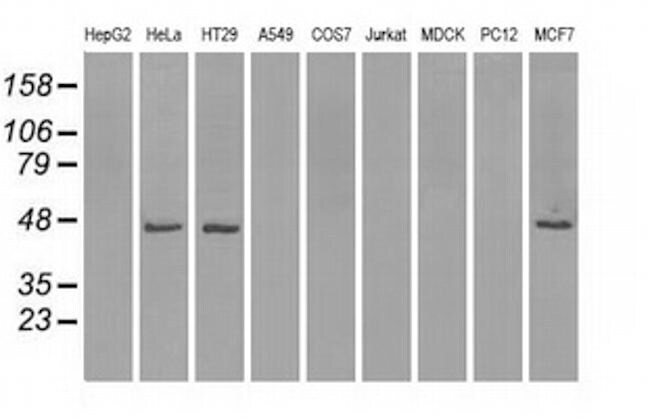
- Western blot analysis of IDH1 in HepG2, HeLa, HT29, A549, COS7, Jurkat, MDCK, PC12, MCF7 cells using 35 µg per lane. Samples were probed with IDH1 (Product # MA5-24966) monoclonal antibody.
- Submitted by
- Invitrogen Antibodies (provider)
- Main image
- Experimental details
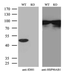
- Western blot analysis of IDH1 in HeLa wild-type and knockout cells using 10 µg per lane. Samples were separated by SDS-PAGE and probed with IDH1 (Product # MA5-24966) monoclonal antibody and HSP90AB1 antibody with a dilution of 1:500 as a loading control.

- Submitted by
- Invitrogen Antibodies (provider)
- Main image
- Experimental details
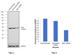
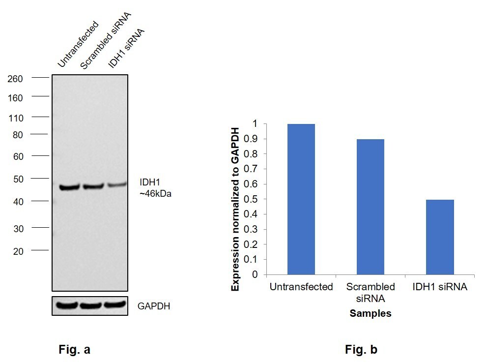
- Knockdown of IDH1 was achieved by transfecting Hep G2 with IDH1 specific siRNAs (Silencer® select Product # S7120, S7121). Western blot analysis (Fig. a) was performed using Whole cell extracts from the IDH1 knockdown cells (lane 3), non-targeting scrambled siRNA transfected cells (lane 2) and untransfected cells (lane 1). The blot was probed with IDH1 Monoclonal Antibody (OTI2H9) (Product # MA5-24966, 1:2000 ) and Goat anti-Mouse IgG (H+L) Superclonal™ Recombinant Secondary Antibody, HRP (Product # A28177, 1:4000). Densitometric analysis of this western blot is shown in histogram (Fig. b). Decrease in signal upon siRNA mediated knock down confirms that antibody is specific to IDH1.
- Submitted by
- Invitrogen Antibodies (provider)
- Main image
- Experimental details
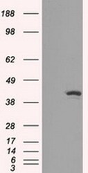
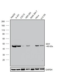
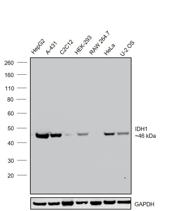
- Western blot was performed using Anti-IDH1 Monoclonal Antibody (OTI2H9) (Product # MA5-24966) and a 46kDa band corresponding to IDH1 was observed across cell lines and tissues tested . Whole cell extracts (30 µg lysate) of Hep G2 (Lane 1), A-431 (Lane 2), C2C12 (Lane 3), HEK-293 (Lane 4), RAW 264.7 (Lane 5), HeLa (Lane 6), U-2 OS (Lane 7) were electrophoresed using NuPAGE™ 4-12% Bis-Tris Protein Gel (Product # NP0322BOX). Resolved proteins were then transferred onto a Nitrocellulose membrane (Product # LC2002) by iBlot® 2 Dry Blotting System (Product # IB21001). The blot was probed with the primary antibody (1:2000) and detected by chemiluminescence with Goat anti-Mouse IgG (H+L) Superclonal™ Recombinant Secondary Antibody, HRP (Product # A28177,1:4000) using the iBright FL 1000 (Product # A32752). Chemiluminescent detection was performed using Novex® ECL Chemiluminescent Substrate Reagent Kit (Product # WP20005).
- Submitted by
- Invitrogen Antibodies (provider)
- Main image

- Experimental details
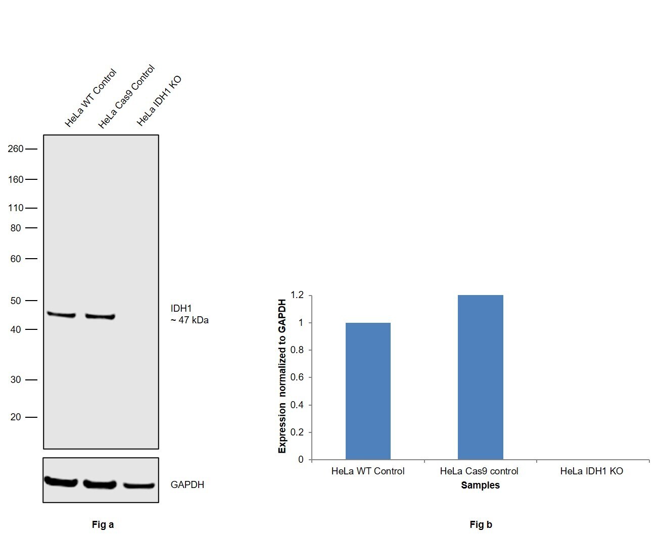
- Knockout of IDH1 was achieved by CRISPR-Cas9 genome editing using LentiArray™ Lentiviral sgRNA (Product # A32042, Assay ID CRISPR1014479_LV) and LentiArray Cas9 Lentivirus (Product # A32064). Western blot analysis of IDH1 was performed by loading 30 µg of HeLa Wild Type (Lane 1), HeLa Cas9 (Lane 2) andHeLa IDH1 KO (Lane 3) whole cell extracts. The samples were electrophoresed using NuPAGE™ Novex™ 4-12% Bis-Tris Protein Gel (Product # NP0322BOX). Resolved proteins were then transferred onto a nitrocellulose membrane (Product # IB23001) by iBlot® 2 Dry Blotting System (Product # IB21001). The blot was probed with an IDH1 Monoclonal Antibody (OTI2H9) (Product # MA5-24966, 1:2,000 dilution) and Goat anti-Mouse IgG (H+L) Superclonal™ Recombinant Secondary Antibody, HRP (Product # A28179, 1:4,000 dilution) using the iBright FL 1000 (Product # A32752). Chemiluminescent detection was performed using Novex® ECL Chemiluminescent Substrate Reagent Kit (Product # WP20005). Loss of signal upon CRISPR mediated knockout (KO) using the LentiArray™ CRISPR product line confirms that antibody is specific to IDH1.
Supportive validation
- Submitted by
- Invitrogen Antibodies (provider)
- Main image
- Experimental details
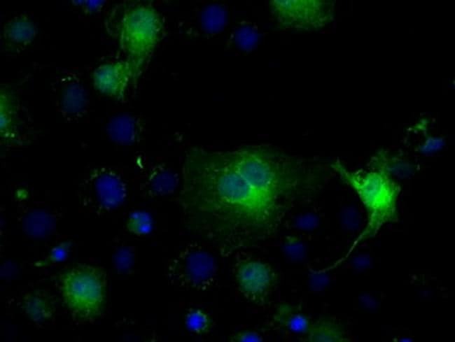
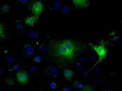
- Immunofluorescent analysis of IDH1 in COS7 cells. Cells were transfected with a plasmid overexpressing IDH1 and probed with a IDH1 monoclonal antibody (Product # MA5-24966).

- Submitted by
- Invitrogen Antibodies (provider)
- Main image

- Experimental details
- Immunofluorescent analysis of IDH1 in COS7 cells. Cells were transfected with a plasmid overexpressing IDH1 and probed with a IDH1 monoclonal antibody (Product # MA5-24966).