Explore
 Validate
 Learn
Learn Western blot
 ELISA
ELISAAntibody data
- Antibody Data
- Antigen structure
- References [0]
- Comments [0]
- Validations
- Western blot [1]
- Immunohistochemistry [16]
Submit
Validation data
Reference
Comment
Report error
- Product number
- STJ98583 - Provider product page

- Provider
- St John's Laboratory
- Product name
- Anti-PTEN antibody (251-300) (STJ98583)
- Antibody type
- Polyclonal
- Description
- Rabbit polyclonal antibody anti-Phosphatidylinositol 3-4-5-Trisphosphate 3-Phosphatase And Dual-Specificity Protein Phosphatase Pten (251-300) is suitable for use in Immunofluorescence, Immunocytochemistry, Western Blot, Immunohistochemistry and ELIS
- Reactivity
- Human, Mouse, Rat
- Host
- Rabbit
- Conjugate
- Unconjugated
- Antigen sequence
NA- Epitope
- NA
- Isotype
- IgG
- Antibody clone number
- NA
- Vial size
- NA
- Concentration
- NA
- Storage
- Store at-20°C for up to 1 year from the date of receipt, and avoid repeat freeze-thaw cycles.
- Handling
- NA
No comments: Submit comment
Supportive validation
- Submitted by
- St John's Laboratory (provider)
- Main image
- Experimental details
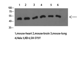
- Western blot analysis of mouse-heart mouse-brain mouse-lung Hela KB SH-SY5Y cells using PTEN Polyclonal Antibody diluted at 1:1000. Secondary antibody was diluted at 1:20000
- Sample type
- NA
- Validation comment
- NA
- Primary Ab dilution
- NA
- Other comments
- NA
- Secondary Ab
- NA
- Secondary Ab dilution
- NA
- Protocol
- NA

Supportive validation
Supportive validation
Supportive validation
Supportive validation
Supportive validation
Supportive validation
Supportive validation
Supportive validation
Supportive validation
Supportive validation
Supportive validation
Supportive validation
Supportive validation
Supportive validation
Supportive validation
Supportive validation
- Submitted by
- St John's Laboratory (provider)
- Main image
- Experimental details
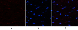
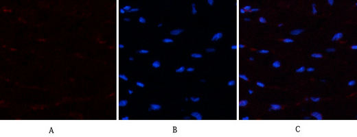
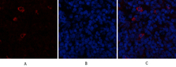
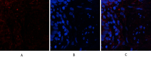
- Immunofluorescence analysis of human-heart tissue. 1, PTEN Polyclonal Antibody (red) was diluted at 1:200 (4°C, overnight). 2, Cy3 labled Secondary antibody was diluted at 1:300 (room temperature, 50min).3, Picture B: DAPI (blue) 10min. Picture A:Target. Picture B: DAPI. Picture C: merge of A+B
- Sample type
- NA
- Validation comment
- NA
- Primary Ab dilution
- NA
- Other comments
- NA
- Secondary Ab
- NA
- Secondary Ab dilution
- NA
- Protocol
- NA
Supportive validation
- Submitted by
- St John's Laboratory (provider)
- Main image
- Experimental details
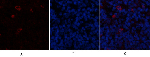
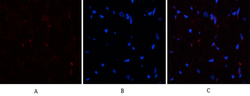
- Immunofluorescence analysis of rat-spleen tissue. 1, PTEN Polyclonal Antibody (red) was diluted at 1:200 (4°C, overnight). 2, Cy3 labled Secondary antibody was diluted at 1:300 (room temperature, 50min).3, Picture B: DAPI (blue) 10min. Picture A:Target. Picture B: DAPI. Picture C: merge of A+B
- Sample type
- NA
- Validation comment
- NA
- Primary Ab dilution
- NA
- Other comments
- NA
- Secondary Ab
- NA
- Secondary Ab dilution
- NA
- Protocol
- NA
Supportive validation
- Submitted by
- St John's Laboratory (provider)
- Main image
- Experimental details
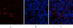
- Immunofluorescence analysis of rat-spleen tissue. 1, PTEN Polyclonal Antibody (red) was diluted at 1:200 (4°C, overnight). 2, Cy3 labled Secondary antibody was diluted at 1:300 (room temperature, 50min).3, Picture B: DAPI (blue) 10min. Picture A:Target. Picture B: DAPI. Picture C: merge of A+B
- Sample type
- NA
- Validation comment
- NA
- Primary Ab dilution
- NA
- Other comments
- NA
- Secondary Ab
- NA
- Secondary Ab dilution
- NA
- Protocol
- NA
Supportive validation
- Submitted by
- St John's Laboratory (provider)
- Main image

- Experimental details
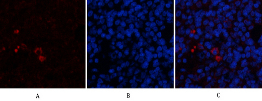
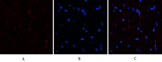
- Immunofluorescence analysis of human-lung tissue. 1, PTEN Polyclonal Antibody (red) was diluted at 1:200 (4°C, overnight). 2, Cy3 labled Secondary antibody was diluted at 1:300 (room temperature, 50min).3, Picture B: DAPI (blue) 10min. Picture A:Target. Picture B: DAPI. Picture C: merge of A+B
- Sample type
- NA
- Validation comment
- NA
- Primary Ab dilution
- NA
- Other comments
- NA
- Secondary Ab
- NA
- Secondary Ab dilution
- NA
- Protocol
- NA
Supportive validation
- Submitted by
- St John's Laboratory (provider)
- Main image

- Experimental details
- Immunofluorescence analysis of human-lung tissue. 1, PTEN Polyclonal Antibody (red) was diluted at 1:200 (4°C, overnight). 2, Cy3 labled Secondary antibody was diluted at 1:300 (room temperature, 50min).3, Picture B: DAPI (blue) 10min. Picture A:Target. Picture B: DAPI. Picture C: merge of A+B
- Sample type
- NA
- Validation comment
- NA
- Primary Ab dilution
- NA
- Other comments
- NA
- Secondary Ab
- NA
- Secondary Ab dilution
- NA
- Protocol
- NA
Supportive validation
- Submitted by
- St John's Laboratory (provider)
- Main image
- Experimental details
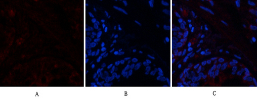
- Immunofluorescence analysis of human-heart tissue. 1, PTEN Polyclonal Antibody (red) was diluted at 1:200 (4°C, overnight). 2, Cy3 labled Secondary antibody was diluted at 1:300 (room temperature, 50min).3, Picture B: DAPI (blue) 10min. Picture A:Target. Picture B: DAPI. Picture C: merge of A+B
- Sample type
- NA
- Validation comment
- NA
- Primary Ab dilution
- NA
- Other comments
- NA
- Secondary Ab
- NA
- Secondary Ab dilution
- NA
- Protocol
- NA
Supportive validation
- Submitted by
- St John's Laboratory (provider)
- Main image
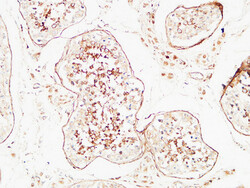
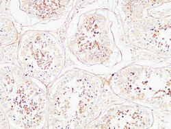
- Experimental details
- Immunohistochemical analysis of paraffin-embedded Human testis. 1, Antibody was diluted at 1:200 (4°C overnight). 2, High-pressure and temperature EDTA, pH8.0 was used for antigen retrieval. 3, Secondary antibody was diluted at 1:200 (room temperature, 30min).
- Sample type
- NA
- Validation comment
- NA
- Primary Ab dilution
- NA
- Other comments
- NA
- Secondary Ab
- NA
- Secondary Ab dilution
- NA
- Protocol
- NA
Supportive validation
- Submitted by
- St John's Laboratory (provider)
- Main image
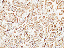
- Experimental details
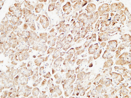
- Immunohistochemical analysis of paraffin-embedded Human pancreas. 1, Antibody was diluted at 1:200 (4°C overnight). 2, High-pressure and temperature EDTA, pH8.0 was used for antigen retrieval. 3, Secondary antibody was diluted at 1:200 (room temperature, 30min).
- Sample type
- NA
- Validation comment
- NA
- Primary Ab dilution
- NA
- Other comments
- NA
- Secondary Ab
- NA
- Secondary Ab dilution
- NA
- Protocol
- NA
Supportive validation
- Submitted by
- St John's Laboratory (provider)
- Main image
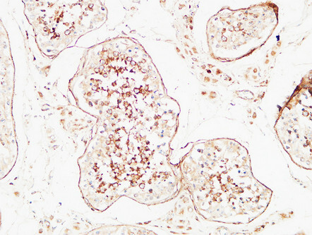
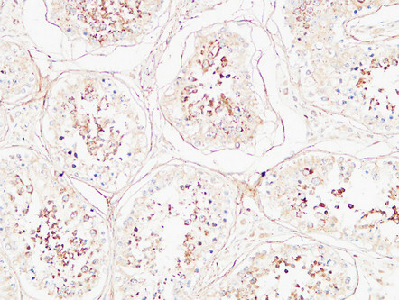
- Experimental details
- Immunohistochemical analysis of paraffin-embedded Human testis. 1, Antibody was diluted at 1:200 (4°C overnight). 2, High-pressure and temperature EDTA, pH8.0 was used for antigen retrieval. 3, Secondary antibody was diluted at 1:200 (room temperature, 30min).
- Sample type
- NA
- Validation comment
- NA
- Primary Ab dilution
- NA
- Other comments
- NA
- Secondary Ab
- NA
- Secondary Ab dilution
- NA
- Protocol
- NA
Supportive validation
- Submitted by
- St John's Laboratory (provider)
- Main image
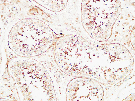
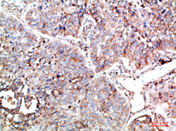
- Experimental details
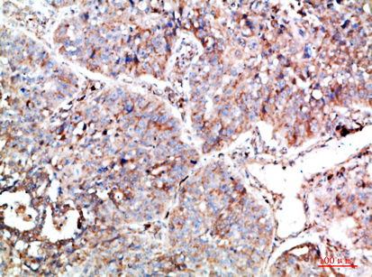
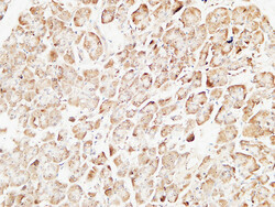
- Immunohistochemical analysis of paraffin-embedded human-lung-cancer, antibody was diluted at 1:200
- Sample type
- NA
- Validation comment
- NA
- Primary Ab dilution
- NA
- Other comments
- NA
- Secondary Ab
- NA
- Secondary Ab dilution
- NA
- Protocol
- NA
Supportive validation
- Submitted by
- St John's Laboratory (provider)
- Main image
- Experimental details
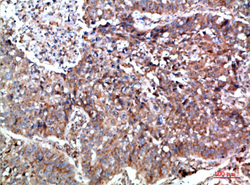
- Immunohistochemical analysis of paraffin-embedded human-lung-cancer, antibody was diluted at 1:200
- Sample type
- NA
- Validation comment
- NA
- Primary Ab dilution
- NA
- Other comments
- NA
- Secondary Ab
- NA
- Secondary Ab dilution
- NA
- Protocol
- NA

Supportive validation
- Submitted by
- St John's Laboratory (provider)
- Main image

- Experimental details
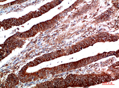
- Immunohistochemical analysis of paraffin-embedded human-colon-cancer, antibody was diluted at 1:200
- Sample type
- NA
- Validation comment
- NA
- Primary Ab dilution
- NA
- Other comments
- NA
- Secondary Ab
- NA
- Secondary Ab dilution
- NA
- Protocol
- NA

Supportive validation
- Submitted by
- St John's Laboratory (provider)
- Main image
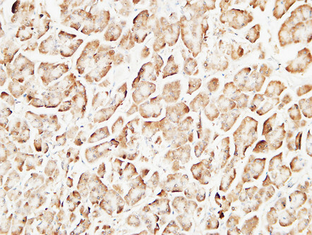
- Experimental details
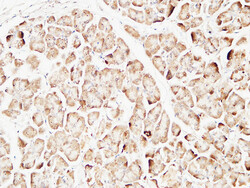
- Immunohistochemical analysis of paraffin-embedded Human pancreas. 1, Antibody was diluted at 1:200 (4°C overnight). 2, High-pressure and temperature EDTA, pH8.0 was used for antigen retrieval. 3, Secondary antibody was diluted at 1:200 (room temperature, 30min).
- Sample type
- NA
- Validation comment
- NA
- Primary Ab dilution
- NA
- Other comments
- NA
- Secondary Ab
- NA
- Secondary Ab dilution
- NA
- Protocol
- NA
Supportive validation
- Submitted by
- St John's Laboratory (provider)
- Main image

- Experimental details
- Immunohistochemical analysis of paraffin-embedded human-colon-cancer, antibody was diluted at 1:200
- Sample type
- NA
- Validation comment
- NA
- Primary Ab dilution
- NA
- Other comments
- NA
- Secondary Ab
- NA
- Secondary Ab dilution
- NA
- Protocol
- NA
Supportive validation
- Submitted by
- St John's Laboratory (provider)
- Main image
- Experimental details
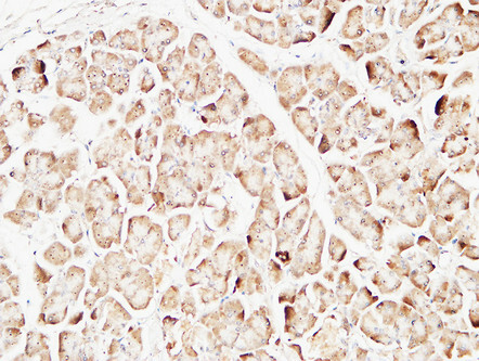
- Immunohistochemical analysis of paraffin-embedded Human pancreas. 1, Antibody was diluted at 1:200 (4°C overnight). 2, High-pressure and temperature EDTA, pH8.0 was used for antigen retrieval. 3, Secondary antibody was diluted at 1:200 (room temperature, 30min).
- Sample type
- NA
- Validation comment
- NA
- Primary Ab dilution
- NA
- Other comments
- NA
- Secondary Ab
- NA
- Secondary Ab dilution
- NA
- Protocol
- NA