Explore
 Validate
 Learn
Learn Western blot
Antibody data
- Antibody Data
- Antigen structure
- References [4]
- Comments [0]
- Validations
- Western blot [2]
- Immunohistochemistry [1]
- Flow cytometry [1]
Submit
Validation data
Reference
Comment
Report error
- Product number
- MAB847 - Provider product page

- Provider
- R&D Systems
- Product name
- Human/Mouse/Rat PTEN Antibody
- Antibody type
- Monoclonal
- Description
- Protein A or G purified from hybridoma culture supernatant. Detects human, mouse, and rat PTEN.
- Reactivity
- Human, Mouse, Rat
- Host
- Mouse
- Conjugate
- Unconjugated
- Antigen sequence
P60484- Isotype
- IgG
- Antibody clone number
- 217702
- Vial size
- 100 ug
- Concentration
- LYOPH
- Storage
- Use a manual defrost freezer and avoid repeated freeze-thaw cycles. 12 months from date of receipt, -20 to -70 °C as supplied. 1 month, 2 to 8 °C under sterile conditions after reconstitution. 6 months, -20 to -70 °C under sterile conditions after reconstitution.
Submitted references Changes in expression of special AT-rich sequence binding protein 1 and phosphatase and tensin homologue in kidneys of diabetic rats during ageing.
MiR221 promotes precursor B-cell retention in the bone marrow by amplifying the PI3K-signaling pathway in mice.
Cancer cell-oriented migration of mesenchymal stem cells engineered with an anticancer gene (PTEN): an imaging demonstration.
Reversal of the malignant phenotype of ovarian cancer A2780 cells through transfection with wild-type PTEN gene.
Delic Jukic IK, Kostic S, Filipovic N, Gudelj Ensor L, Ivandic M, Dukic JJ, Vitlov Uljevic M, Ferhatovic Hamzic L, Puljak L, Vukojevic K
Nephrology, dialysis, transplantation : official publication of the European Dialysis and Transplant Association - European Renal Association 2018 Oct 1;33(10):1734-1741
Nephrology, dialysis, transplantation : official publication of the European Dialysis and Transplant Association - European Renal Association 2018 Oct 1;33(10):1734-1741
MiR221 promotes precursor B-cell retention in the bone marrow by amplifying the PI3K-signaling pathway in mice.
Petkau G, Kawano Y, Wolf I, Knoll M, Melchers F
European journal of immunology 2018 Jun;48(6):975-989
European journal of immunology 2018 Jun;48(6):975-989
Cancer cell-oriented migration of mesenchymal stem cells engineered with an anticancer gene (PTEN): an imaging demonstration.
Yang ZS, Tang XJ, Guo XR, Zou DD, Sun XY, Feng JB, Luo J, Dai LJ, Warnock GL
OncoTargets and therapy 2014;7:441-6
OncoTargets and therapy 2014;7:441-6
Reversal of the malignant phenotype of ovarian cancer A2780 cells through transfection with wild-type PTEN gene.
Wu H, Wang S, Weng D, Xing H, Song X, Zhu T, Xia X, Weng Y, Xu G, Meng L, Zhou J, Ma D
Cancer letters 2008 Nov 28;271(2):205-14
Cancer letters 2008 Nov 28;271(2):205-14
No comments: Submit comment
Supportive validation
- Submitted by
- R&D Systems (provider)
- Main image
- Experimental details
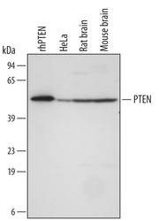
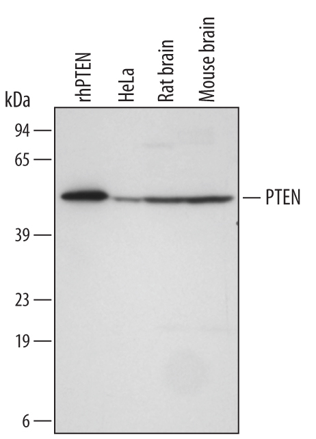
- Detection of Human/Mouse/Rat PTEN by Western Blot. Western blot shows lysates of HeLa human cervical epithelial carcinoma cell line and rat and mouse brain tissue. PVDF membrane was probed with 0.5 µg/mL Mouse Anti-Human/Mouse/Rat PTEN Monoclonal Antibody (Catalog # MAB847) followed by HRP-conjugated Anti-Mouse IgG Secondary Antibody (Catalog # HAF007). For additional reference, Recombinant Human PTEN (Catalog # 847-PN) (5 ng) was included. A specific band for PTEN was detected at approximately 54 kDa (as indicated). This experiment was conducted under reducing conditions and using Immunoblot Buffer Group 4.
- Submitted by
- R&D Systems (provider)
- Main image

- Experimental details
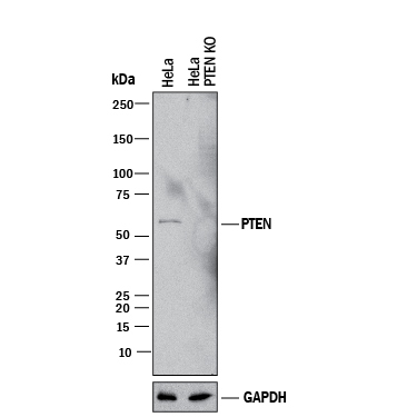
- Western Blot Shows Human PTEN Specificity by Using Knockout Cell Line. Western blot shows lysates of HeLa human cervical epithelial carcinoma parental cell line and PTEN knockout HeLa cell line (KO). PVDF membrane was probed with 0.5 µg/mL of Mouse Anti-Human/Mouse/Rat PTEN Monoclonal Antibody (Catalog # MAB847) followed by HRP-conjugated Anti-Mouse IgG Secondary Antibody (Catalog # HAF018). A specific band was detected for PTEN at approximately 55 kDa (as indicated) in the parental HeLa cell line, but is not detectable in knockout HeLa cell line. GAPDH (Catalog # MAB5718) is shown as a loading control. This experiment was conducted under reducing conditions and using Immunoblot Buffer Group 1.
Supportive validation
- Submitted by
- R&D Systems (provider)
- Main image
- Experimental details
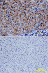
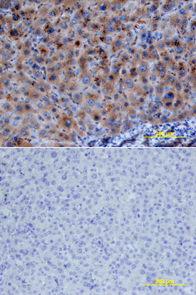
- PTEN in Human Liver. PTEN was detected in immersion fixed paraffin-embedded sections of human liver array using Mouse Anti-Human/Mouse/Rat PTEN Monoclonal Antibody (Catalog # MAB847) at 25 µg/mL overnight at 4 °C. Tissue was stained using the Anti-Mouse HRP-DAB Cell & Tissue Staining Kit (brown; Catalog # CTS002) and counterstained with hematoxylin (blue). Lower panel shows a lack of labeling if primary antibodies are omitted and tissue is stained only with secondary antibody followed by incubation with detection reagents. View our protocol for Chromogenic IHC Staining of Paraffin-embedded Tissue Sections.
Supportive validation
- Submitted by
- R&D Systems (provider)
- Main image
- Experimental details
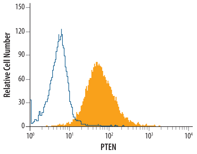
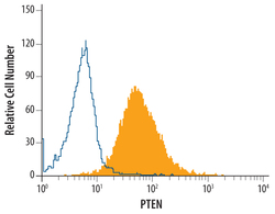
- Detection of PTEN in Human PBMC lymphocytes by Flow Cytometry. Human peripheral blood mononuclear cell lymphocytes were stained with Mouse Anti-Human/Mouse/Rat PTEN Monoclonal Antibody (Catalog # MAB847, filled histogram) or isotype control antibody (Catalog # MAB002, open histogram), followed by Phycoerythrin-conjugated Anti-Mouse IgG F(ab')2 Secondary Antibody (Catalog # F0102B). To facilitate intracellular staining, cells were fixed with paraformaldehyde and permeabilized with saponin.