 Explore
 Validate
 Learn
Learn Western blot
 Immunocytochemistry
Antibody data
- Antibody Data
- Antigen structure
- References [2]
- Comments [0]
- Validations
- Immunocytochemistry [4]
- Immunoprecipitation [2]
- Other assay [6]
Submit
Validation data
Reference
Comment
Report error
- Product number
- MA1-089 - Provider product page

- Provider
- Invitrogen Antibodies
- Product name
- eIF4E Monoclonal Antibody (5D11)
- Antibody type
- Monoclonal
- Antigen
- Other
- Description
- MA1-089 detects eIF4E in human and mouse samples and has been successfully used in Western blot, immunofluorescence, immunocytochemistry and immunoprecipitation applications. MA1-089 does not react with rat eIF4E. The MA1-089 immunogen is full length human eIF4E produced in HEK293T cells.
- Reactivity
- Human, Mouse
- Host
- Mouse
- Isotype
- IgG
- Antibody clone number
- 5D11
- Vial size
- 100 μg
- Concentration
- 1 mg/mL
- Storage
- -20°C
Submitted references mTOR-dependent translation amplifies microglia priming in aging mice.
Nuclear localization of EIF4G3 suggests a role for the XY body in translational regulation during spermatogenesis in mice.
Keane L, Antignano I, Riechers SP, Zollinger R, Dumas AA, Offermann N, Bernis ME, Russ J, Graelmann F, McCormick PN, Esser J, Tejera D, Nagano A, Wang J, Chelala C, Biederbick Y, Halle A, Salomoni P, Heneka MT, Capasso M
The Journal of clinical investigation 2021 Jan 4;131(1)
The Journal of clinical investigation 2021 Jan 4;131(1)
Nuclear localization of EIF4G3 suggests a role for the XY body in translational regulation during spermatogenesis in mice.
Hu J, Sun F, Handel MA
Biology of reproduction 2018 Jan 1;98(1):102-114
Biology of reproduction 2018 Jan 1;98(1):102-114
No comments: Submit comment
Supportive validation
- Submitted by
- Invitrogen Antibodies (provider)
- Main image
- Experimental details
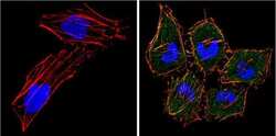
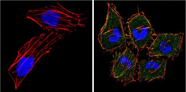
- Immunofluorescent analysis of eIF4E (green) showing positive staining in the cytoplasm of Hela cells (right) compared with a negative control in the absence of primary antibody (left). Formalin-fixed cells were permeabilized with 0.1% Triton X-100 in TBS for 5-10 minutes, blocked with 3% BSA-PBS for 30 minutes at room temperature and probed with an eIF4E monoclonal antibody (Product # MA1-089) in 3% BSA-PBS at a dilution of 1:100 and incubated overnight at 4 °C in a humidified chamber. Cells were washed with PBST and incubated with a DyLight 488-conjugated goat-anti-mouse IgG (H+L) secondary antibody in PBS at room temperature in the dark. F-actin (red) was stained with a fluorescent red phalloidin and nuclei (blue) were stained with DAPI for 5-10 minutes in the dark. Images were taken at a magnification of 60x.
- Submitted by
- Invitrogen Antibodies (provider)
- Main image
- Experimental details
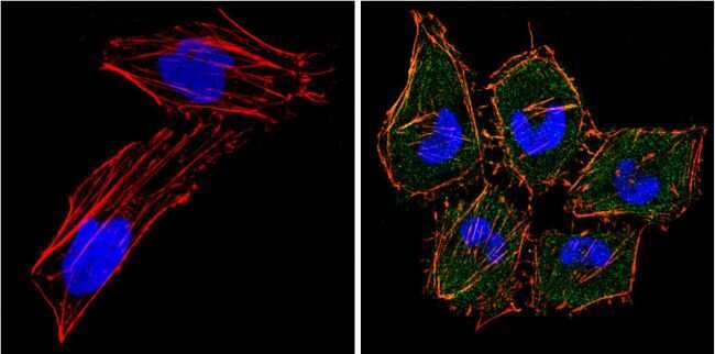
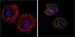
- Immunofluorescent analysis of eIF4E (green) showing positive staining in the cytoplasm of MCF-7 cells (right) compared with a negative control in the absence of primary antibody (left). Formalin-fixed cells were permeabilized with 0.1% Triton X-100 in TBS for 5-10 minutes, blocked with 3% BSA-PBS for 30 minutes at room temperature and probed with an eIF4E monoclonal antibody (Product # MA1-089) in 3% BSA-PBS at a dilution of 1:100 and incubated overnight at 4 °C in a humidified chamber. Cells were washed with PBST and incubated with a DyLight 488-conjugated goat-anti-mouse IgG (H+L) secondary antibody in PBS at room temperature in the dark. F-actin (red) was stained with a fluorescent red phalloidin and nuclei (blue) were stained with DAPI for 5-10 minutes in the dark. Images were taken at a magnification of 60x.
- Submitted by
- Invitrogen Antibodies (provider)
- Main image

- Experimental details
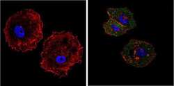
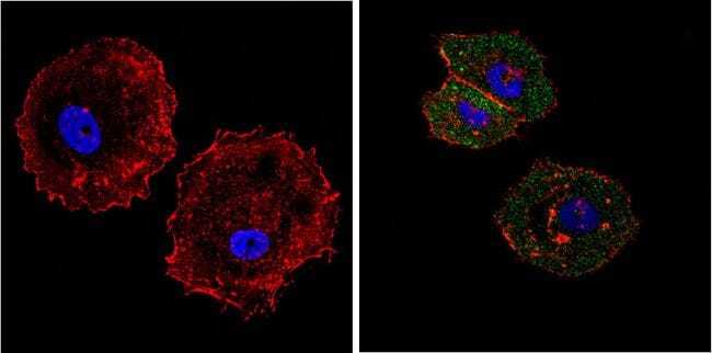
- Immunofluorescent analysis of eIF4E (green) showing positive staining in the cytoplasm of Hela cells (right) compared with a negative control in the absence of primary antibody (left). Formalin-fixed cells were permeabilized with 0.1% Triton X-100 in TBS for 5-10 minutes, blocked with 3% BSA-PBS for 30 minutes at room temperature and probed with an eIF4E monoclonal antibody (Product # MA1-089) in 3% BSA-PBS at a dilution of 1:100 and incubated overnight at 4 °C in a humidified chamber. Cells were washed with PBST and incubated with a DyLight 488-conjugated goat-anti-mouse IgG (H+L) secondary antibody in PBS at room temperature in the dark. F-actin (red) was stained with a fluorescent red phalloidin and nuclei (blue) were stained with DAPI for 5-10 minutes in the dark. Images were taken at a magnification of 60x.
- Submitted by
- Invitrogen Antibodies (provider)
- Main image
- Experimental details
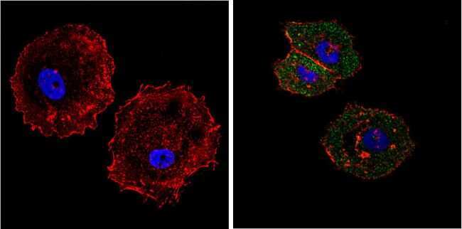
- Immunofluorescent analysis of eIF4E (green) showing positive staining in the cytoplasm of MCF-7 cells (right) compared with a negative control in the absence of primary antibody (left). Formalin-fixed cells were permeabilized with 0.1% Triton X-100 in TBS for 5-10 minutes, blocked with 3% BSA-PBS for 30 minutes at room temperature and probed with an eIF4E monoclonal antibody (Product # MA1-089) in 3% BSA-PBS at a dilution of 1:100 and incubated overnight at 4 °C in a humidified chamber. Cells were washed with PBST and incubated with a DyLight 488-conjugated goat-anti-mouse IgG (H+L) secondary antibody in PBS at room temperature in the dark. F-actin (red) was stained with a fluorescent red phalloidin and nuclei (blue) were stained with DAPI for 5-10 minutes in the dark. Images were taken at a magnification of 60x.
Supportive validation
- Submitted by
- Invitrogen Antibodies (provider)
- Main image
- Experimental details
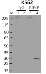
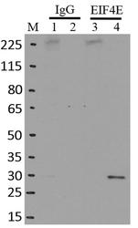
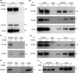
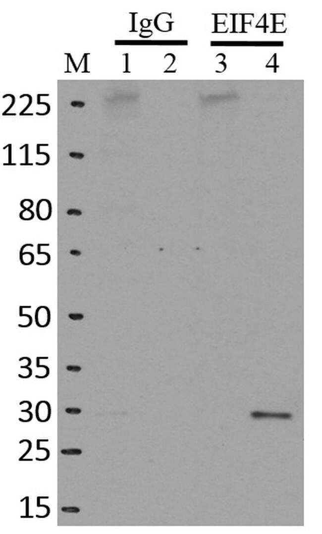
- Immunoprecipitation of EIF4E was performed on K562 cells. Antigen-antibody complexes were formed by incubating approximately 500 µg whole cell lysate with 5 µg of monoclonal EIF4E antibody (Product # MA1-089) rotating 60 min at RT. The immune complexes were captured on 625 µg of anti-mouse coated Dynabeads (Product # 11202D), washed extensively, and eluted with NuPAGE™ LDS Sample Buffer (Product # NP0007). Samples were resolved onto NuPAGE™ 4-12% Bis-Tris gel (Product # NP0335BOX). Lanes 1 and 3 are input and lanes 2 and 4 are IP. Proteins were transferred to PVDF membrane (Product # IB23001). Membrane was blocked in 5% milk. Target was detected using a EIF4E monoclonal antibody (Product # MA1-089) at a dilution of 1:2,000, followed by a 1:4,000 dilution of secondary antibody. Chemiluminescent detection was performed using ECL Western Blotting Substrate (Product # 32106). Data courtesy of the Yeo lab as part of the ENCODE project (www.encodeproject.org).

- Submitted by
- Invitrogen Antibodies (provider)
- Main image

- Experimental details
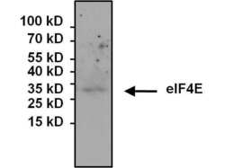
- Immunoprecipitation of eIF4E was performed on HeLa cell lysates. The antigen:antibody complex was formed by incubating 500 µg whole cell lysate with 3 µg of mouse monoclonal antibody recognizing eIF4E (Product # MA1-089) overnight on a rocking platform at 4°C. The immune-complex was captured on 50 µL Protein A/G Plus Agarose (Product # 20423). Captured immune-complexes were then washed extensively and proteins eluted with 5X Reducing Sample Loading Dye (Product # 39000). Samples were resolved on a 4-20% Tris-HCl polyacrylamide gel. Proteins were transferred to PVDF membrane and blocked with 5% Milk/TBS-0.1%Tween for at least 1 hour. Membranes were then probed with a mouse monoclonal antibody recognizing eIF4E (Product # MA1-089) at a dilution of 1:1000 overnight at 4°C on a rocking platform. Membranes were washed in TBST and probed with Pierce Clean Blot (Product # 21230) at a dilution of 1:1,000 for at least one hour. Membranes were washed and chemiluminescent detection performed using Super Signal West Dura (Product # 34075).

Supportive validation
- Submitted by
- Invitrogen Antibodies (provider)
- Main image
- Experimental details
- RNA immunoprecipitation (RIP) western of EIF4E was performed on K562 cells. Antigen-antibody complexes were formed by incubating approximately 500 µg whole cell lysate with 5 µg of monoclonal EIF4E antibody (Product # MA1-089) rotating 60 min at RT. The immune complexes were captured on 625 µg of anti-mouse coated Dynabeads (Product # 11202D), washed extensively, and eluted with NuPAGE™ LDS Sample Buffer (Product # NP0007). Samples were resolved onto NuPAGE™ 4-12% Bis-Tris gel (Product # NP0335BOX). Lanes 1 and 3 are input and lanes 2 and 4 are IP. Proteins were transferred to PVDF membrane (Product # IB23001). Membrane was blocked in 5% milk. Target was detected using a EIF4E monoclonal antibody (Product # MA1-089) at a dilution of 1:2,000, followed by a 1:4000 dilution of secondary antibody. Chemiluminescent detection was performed using ECL Western Blotting Substrate (Product # 32106). Data courtesy of the Yeo lab as part of the ENCODE project (www.encodeproject.org).

- Submitted by
- Invitrogen Antibodies (provider)
- Main image
- Experimental details
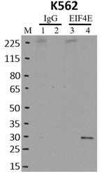
- Immunoprecipitation of EIF4E was performed on K562 cells. Antigen-antibody complexes were formed by incubating approximately 500 µg whole cell lysate with 5 µg of monoclonal EIF4E antibody (Product # MA1-089) rotating 60 min at RT. The immune complexes were captured on 625 µg of anti-mouse coated Dynabeads (Product # 11202D), washed extensively, and eluted with NuPAGE™ LDS Sample Buffer (Product # NP0007). Samples were resolved onto NuPAGE™ 4-12% Bis-Tris gel (Product # NP0335BOX). Lanes 1 and 3 are input and lanes 2 and 4 are IP. Proteins were transferred to PVDF membrane (Product # IB23001). Membrane was blocked in 5% milk. Target was detected using a EIF4E monoclonal antibody (Product # MA1-089) at a dilution of 1:2,000, followed by a 1:4,000 dilution of secondary antibody. Chemiluminescent detection was performed using ECL Western Blotting Substrate (Product # 32106). Data courtesy of the Yeo lab as part of the ENCODE project (www.encodeproject.org).

- Submitted by
- Invitrogen Antibodies (provider)
- Main image
- Experimental details
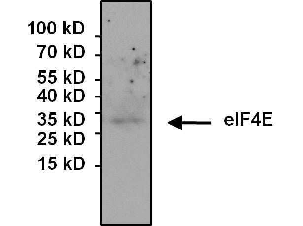
- Immunoprecipitation of eIF4E was performed on HeLa cell lysates. The antigen:antibody complex was formed by incubating 500 µg whole cell lysate with 3 µg of mouse monoclonal antibody recognizing eIF4E (Product # MA1-089) overnight on a rocking platform at 4øC. The immune-complex was captured on 50 µL Protein A/G Plus Agarose (Product # 20423). Captured immune-complexes were then washed extensively and proteins eluted with 5X Reducing Sample Loading Dye (Product # 39000). Samples were resolved on a 4-20% Tris-HCl polyacrylamide gel. Proteins were transferred to PVDF membrane and blocked with 5% Milk/TBS-0.1%Tween for at least 1 hour. Membranes were then probed with a mouse monoclonal antibody recognizing eIF4E (Product # MA1-089) at a dilution of 1:1000 overnight at 4øC on a rocking platform. Membranes were washed in TBST and probed with Pierce Clean Blot (Product # 21230) at a dilution of 1:1,000 for at least one hour. Membranes were washed and chemiluminescent detection performed using Super Signal West Dura (Product # 34075).
- Submitted by
- Invitrogen Antibodies (provider)
- Main image
- Experimental details
- NULL

- Submitted by
- Invitrogen Antibodies (provider)
- Main image

- Experimental details
- RNA immunoprecipitation (RIP) western of EIF4E was performed on K562 cells. Antigen-antibody complexes were formed by incubating approximately 500 µg whole cell lysate with 5 µg of monoclonal EIF4E antibody (Product # MA1-089) rotating 60 min at RT. The immune complexes were captured on 625 µg of anti-mouse coated Dynabeads (Product # 11202D), washed extensively, and eluted with NuPAGE™ LDS Sample Buffer (Product # NP0007). Samples were resolved onto NuPAGE™ 4-12% Bis-Tris gel (Product # NP0335BOX). Lanes 1 and 3 are input and lanes 2 and 4 are IP. Proteins were transferred to PVDF membrane (Product # IB23001). Membrane was blocked in 5% milk. Target was detected using a EIF4E monoclonal antibody (Product # MA1-089) at a dilution of 1:2,000, followed by a 1:4000 dilution of secondary antibody. Chemiluminescent detection was performed using ECL Western Blotting Substrate (Product # 32106). Data courtesy of the Yeo lab as part of the ENCODE project (www.encodeproject.org).
- Submitted by
- Invitrogen Antibodies (provider)
- Main image

- Experimental details
- Immunoprecipitation of EIF4E was performed on K562 cells. Antigen-antibody complexes were formed by incubating approximately 500 µg whole cell lysate with 5 µg of monoclonal EIF4E antibody (Product # MA1-089) rotating 60 min at RT. The immune complexes were captured on 625 µg of anti-mouse coated Dynabeads (Product # 11202D), washed extensively, and eluted with NuPAGE™ LDS Sample Buffer (Product # NP0007). Samples were resolved onto NuPAGE™ 4-12% Bis-Tris gel (Product # NP0335BOX). Lanes 1 and 3 are input and lanes 2 and 4 are IP. Proteins were transferred to PVDF membrane (Product # IB23001). Membrane was blocked in 5% milk. Target was detected using a EIF4E monoclonal antibody (Product # MA1-089) at a dilution of 1:2,000, followed by a 1:4,000 dilution of secondary antibody. Chemiluminescent detection was performed using ECL Western Blotting Substrate (Product # 32106). Data courtesy of the Yeo lab as part of the ENCODE project (www.encodeproject.org).
