Explore
 Validate
 Learn
Learn Western blot
Antibody data
- Antibody Data
- Antigen structure
- References [1]
- Comments [0]
- Validations
- Western blot [2]
- Immunocytochemistry [1]
Submit
Validation data
Reference
Comment
Report error
- Product number
- MAB9027 - Provider product page

- Provider
- R&D Systems
- Product name
- Human/Mouse Phospho-PDGF R beta (Y751) Antibody
- Antibody type
- Monoclonal
- Description
- Protein A or G purified from cell culture supernatant. Detects human and mouse PDGF R beta when phosphorylated at Y751 in Western blots.
- Reactivity
- Human, Mouse
- Host
- Rabbit
- Conjugate
- Unconjugated
- Antigen sequence
P09619- Isotype
- IgG
- Antibody clone number
- 1210B
- Vial size
- 100 ug
- Storage
- Use a manual defrost freezer and avoid repeated freeze-thaw cycles. 12 months from date of receipt, -20 to -70 °C as supplied. 1 month, 2 to 8 °C under sterile conditions after reconstitution. 6 months, -20 to -70 °C under sterile conditions after reconstitution.
Submitted references Assembly of fibronectin fibrils selectively attenuates platelet-derived growth factor-induced intracellular calcium release in fibroblasts.
Farrar CS, Hocking DC
The Journal of biological chemistry 2018 Nov 30;293(48):18655-18666
The Journal of biological chemistry 2018 Nov 30;293(48):18655-18666
No comments: Submit comment
Supportive validation
- Submitted by
- R&D Systems (provider)
- Main image

- Experimental details
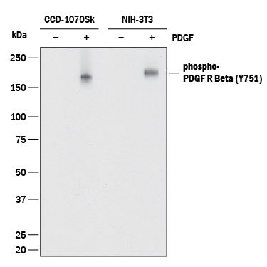
- Detection of Human and Mouse Phospho-PDGF R beta (Y751) by Western Blot. Western blot shows lysates of CCD-1070Sk human foreskin fibroblast cell line and NIH-3T3 mouse embryonic fibroblast cell line untreated (-) or treated (+) with 100 ng/mL Recombinant Human PDGF-BB (Catalog # 220-BB) for 20 minutes. PVDF membrane was probed with 0.1 µg/mL of Rabbit Anti-Human/Mouse Phospho-PDGF R beta (Y751) Monoclonal Antibody (Catalog # MAB9027) followed by HRP-conjugated Anti-Rabbit IgG Secondary Antibody (Catalog # HAF008). A specific band was detected for PDGF R beta at approximately 190 kDa (as indicated). This experiment was conducted under reducing conditions and using Immunoblot Buffer Group 1.
- Submitted by
- R&D Systems (provider)
- Main image

- Experimental details
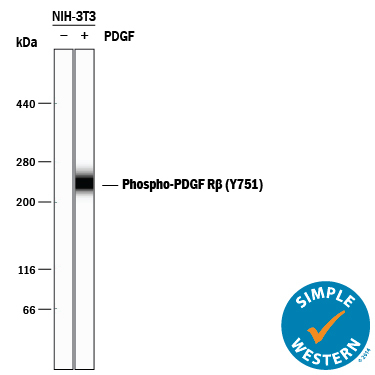
- Detection of Mouse Phospho-PDGF R beta (Y751) by Simple WesternTM. Simple Western lane view shows lysates of NIH-3T3 mouse embryonic fibroblast cell line untreated (-) or treated (+) with 100 ng/mL Recombinant Human PDGF-BB (Catalog # 220-BB) for 20 minutes, loaded at 0.2 mg/mL. A specific band was detected for PDGF R beta at approximately 240 kDa (as indicated) using 1 µg/mL of Rabbit Anti-Human/Mouse Phospho-PDGF R beta (Y751) Monoclonal Antibody (Catalog # MAB9027). This experiment was conducted under reducing conditions and using the 66-440 kDa separation system.
Supportive validation
- Submitted by
- R&D Systems (provider)
- Main image

- Experimental details
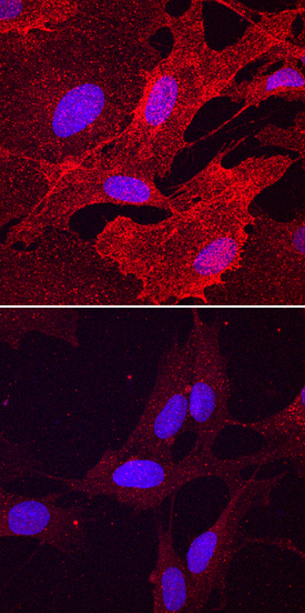
- Phospho-PDGF R beta (Y751) in BJ Human Cell Line. PDGF R beta phosphorylated at Y751 was detected in immersion fixed BJ human skin fibroblast cell line stimulated (top panel), and unstimulated (bottom panel), with Recombinant Human PDGF-BB (Catalog # 220-BB) using Rabbit Anti-Human/Mouse Phospho-PDGF R beta (Y751) Monoclonal Antibody (Catalog # MAB9027) at 5 µg/mL for 3 hours at room temperature. Cells were stained using the NorthernLights™ 557-conjugated Anti-Rabbit IgG Secondary Antibody (red; Catalog # NL004) and counterstained with DAPI (blue). Specific staining was localized to cell surfaces and cytoplasm. View our protocol for Fluorescent ICC Staining of Cells on Coverslips.