 Explore
 Validate
 Learn
Learn Western blot
 Immunoprecipitation
Antibody data
- Antibody Data
- Antigen structure
- References [1]
- Comments [0]
- Validations
- Western blot [1]
- Other assay [1]
Submit
Validation data
Reference
Comment
Report error
- Product number
- MA5-15103 - Provider product page

- Provider
- Invitrogen Antibodies
- Product name
- PDGFRB Monoclonal Antibody (K.596.7)
- Antibody type
- Monoclonal
- Antigen
- Recombinant full-length protein
- Description
- It is not recommended to aliquot this antibody. This antibody is not cross-reactive with PDGF receptor -alpha.
- Reactivity
- Human, Mouse, Rat
- Host
- Mouse
- Isotype
- IgG
- Antibody clone number
- K.596.7
- Vial size
- 100 µL
- Concentration
- 1.75 mg/mL
- Storage
- -20°C
Submitted references Simultaneously targeting cancer-associated fibroblasts and angiogenic vessel as a treatment for TNBC.
Sharma M, Turaga RC, Yuan Y, Satyanarayana G, Mishra F, Bian Z, Liu W, Sun L, Yang J, Liu ZR
The Journal of experimental medicine 2021 Apr 5;218(4)
The Journal of experimental medicine 2021 Apr 5;218(4)
No comments: Submit comment
Supportive validation
- Submitted by
- Invitrogen Antibodies (provider)
- Main image
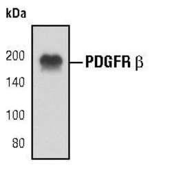
- Experimental details
- Western blot analysis of PDGF Receptor beta in NIH-3T3 cell lysates using PDGF Receptor beta monoclonal antibody (Product # MA5-15103).
Supportive validation
- Submitted by
- Invitrogen Antibodies (provider)
- Main image
- Experimental details
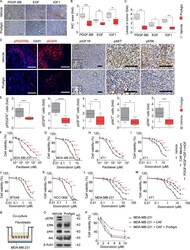
- Figure S3. ProAgio reduces levels of GFs and signaling downstream; the addition of GFs or CAF-conditioned media mediates apoptotic resistance in cancer cells; and ProAgio induces apoptosis in breast CAFs and therefore sensitizes cancer cells to apoptotic induction by a chemotherapeutic agent. (A and B) Representative images of IHC staining of PDGF-BB, EGF, and IGF1 (A) and quantitative analysis of PDGF-BB, EGF, and IGF1-positive area (B) in the tumor sections of 4T1 mice treated with vehicle or ProAgio. Scale bars, 100 um; n = 3-4/group. (C) Intratumoral levels of PDGF-BB, EGF, and IGF1 were determined by ELISA assay in the tumor extracts of 4T1 mice ( n = 4/group). ( D and E ) Representative images of IF staining of pPDGFRbeta, pEGFR, IHC staining of pIGF1R, pAKT, and pERK (D), and quantitative analysis of pPDGFRbeta, pEGFR, pIGF1R, pAKT, and pERK-positive cells (E) in the tumor sections of 4T1 mice treated with vehicle or ProAgio. In IF images, nuclei were counterstained with DAPI (blue). The quantifications are presented as fold change using the ProAgio treatment group as a reference. Scale bars, 100 um; n = 4-5/group. (F-K) Cell viability of TNBC cells, including MDA-MB-231 (F and G), 4T1 (H and I), BT549 (J), and HCC1806 (K) upon treatment with indicated concentrations of indicated drugs for 48 h in the culture media containing the vehicle (i.e., PBS [black line] or indicated GFs [red line]). (L and M) Cell viability of MDA-MB-231 (L) and 4T1 (M) cells upon treatment wit
