Explore
 Validate
 Learn
Learn Western blot
Antibody data
- Antibody Data
- Antigen structure
- References [18]
- Comments [0]
- Validations
- Western blot [1]
- Immunocytochemistry [2]
- Flow cytometry [3]
- Other assay [2]
Submit
Validation data
Reference
Comment
Report error
- Product number
- MA5-13008 - Provider product page

- Provider
- Invitrogen Antibodies
- Product name
- ErbB3 Monoclonal Antibody (H3.105.5 (Ab105))
- Antibody type
- Monoclonal
- Antigen
- Recombinant full-length protein
- Description
- MA5-13008 targets HER-3 in WB, flow cytometry, ICC/IF, and IP applications and shows reactivity with Human and mouse samples.
- Antibody clone number
- H3.105.5 (Ab105)
- Concentration
- 0.2 mg/mL
Submitted references ErbB1-dependent signalling and vesicular trafficking in primary afferent nociceptors associated with hypersensitivity in neuropathic pain.
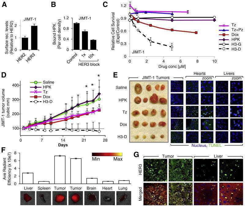
Resistance to receptor-blocking therapies primes tumors as targets for HER3-homing nanobiologics.
Synergistic Chemoimmunotherapy of Epithelial Ovarian Cancer Using ErbB-Retargeted T Cells Combined with Carboplatin.
A Notch1-neuregulin1 autocrine signaling loop contributes to melanoma growth.
Flexible targeting of ErbB dimers that drive tumorigenesis by using genetically engineered T cells.
Decorin, erythroblastic leukaemia viral oncogene homologue B4 and signal transducer and activator of transcription 3 regulation of semaphorin 3A in central nervous system scar tissue.
Re-epithelialization from human skin explant cultures is promoted by ligand-activated HER3 receptor.
Activation of Stat3 by heregulin/ErbB-2 through the co-option of progesterone receptor signaling drives breast cancer growth.
The EGF/CSF-1 paracrine invasion loop can be triggered by heregulin beta1 and CXCL12.
ErbB4 regulates fetal surfactant phospholipid synthesis in primary fetal rat type II cells.
Muc4-ErbB2 complex formation and signaling in polarized CACO-2 epithelial cells indicate that Muc4 acts as an unorthodox ligand for ErbB2.
The role of HER1-HER4 and EGFRvIII in hormone-refractory prostate cancer.
ErbB receptor dimerization, localization, and co-localization in mouse lung type II epithelial cells.
Differential localization of ErbB receptor ensembles influences their signaling in hippocampal neurons.
Signaling pathways required for matrix metalloproteinase-9 induction by betacellulin in head-and-neck squamous carcinoma cells.
Neuregulin expression, function, and signaling in human ovarian cancer cells.
Absence of endothelial cells, central necrosis, and fibrosis are associated with aggressive inflammatory breast cancer.
Epidermal growth factor-like ligands differentially up-regulate matrix metalloproteinase 9 in head and neck squamous carcinoma cells.
Mitchell R, Mikolajczak M, Kersten C, Fleetwood-Walker S
Neurobiology of disease 2020 Aug;142:104961
Neurobiology of disease 2020 Aug;142:104961
Resistance to receptor-blocking therapies primes tumors as targets for HER3-homing nanobiologics.
Sims JD, Taguiam JM, Alonso-Valenteen F, Markman J, Agadjanian H, Chu D, Lubow J, Abrol R, Srinivas D, Jain A, Han B, Qu Y, Mirzadehgan P, Hwang JY, Rentsendorj A, Chung A, Lester J, Karlan BY, Gray HB, Gross Z, Giuliano A, Cui X, Medina-Kauwe LK
Journal of controlled release : official journal of the Controlled Release Society 2018 Feb 10;271:127-138
Journal of controlled release : official journal of the Controlled Release Society 2018 Feb 10;271:127-138
Synergistic Chemoimmunotherapy of Epithelial Ovarian Cancer Using ErbB-Retargeted T Cells Combined with Carboplatin.
Parente-Pereira AC, Whilding LM, Brewig N, van der Stegen SJ, Davies DM, Wilkie S, van Schalkwyk MC, Ghaem-Maghami S, Maher J
Journal of immunology (Baltimore, Md. : 1950) 2013 Sep 1;191(5):2437-45
Journal of immunology (Baltimore, Md. : 1950) 2013 Sep 1;191(5):2437-45
A Notch1-neuregulin1 autocrine signaling loop contributes to melanoma growth.
Zhang K, Wong P, Zhang L, Jacobs B, Borden EC, Aster JC, Bedogni B
Oncogene 2012 Oct 25;31(43):4609-18
Oncogene 2012 Oct 25;31(43):4609-18
Flexible targeting of ErbB dimers that drive tumorigenesis by using genetically engineered T cells.
Davies DM, Foster J, Van Der Stegen SJ, Parente-Pereira AC, Chiapero-Stanke L, Delinassios GJ, Burbridge SE, Kao V, Liu Z, Bosshard-Carter L, Van Schalkwyk MC, Box C, Eccles SA, Mather SJ, Wilkie S, Maher J
Molecular medicine (Cambridge, Mass.) 2012 May 9;18(1):565-76
Molecular medicine (Cambridge, Mass.) 2012 May 9;18(1):565-76
Decorin, erythroblastic leukaemia viral oncogene homologue B4 and signal transducer and activator of transcription 3 regulation of semaphorin 3A in central nervous system scar tissue.
Minor KH, Bournat JC, Toscano N, Giger RJ, Davies SJ
Brain : a journal of neurology 2011 Apr;134(Pt 4):1140-55
Brain : a journal of neurology 2011 Apr;134(Pt 4):1140-55
Re-epithelialization from human skin explant cultures is promoted by ligand-activated HER3 receptor.
Forsberg S, Rollman O
Journal of dermatological science 2010 Jul;59(1):7-15
Journal of dermatological science 2010 Jul;59(1):7-15
Activation of Stat3 by heregulin/ErbB-2 through the co-option of progesterone receptor signaling drives breast cancer growth.
Proietti CJ, Rosemblit C, Beguelin W, Rivas MA, Díaz Flaqué MC, Charreau EH, Schillaci R, Elizalde PV
Molecular and cellular biology 2009 Mar;29(5):1249-65
Molecular and cellular biology 2009 Mar;29(5):1249-65
The EGF/CSF-1 paracrine invasion loop can be triggered by heregulin beta1 and CXCL12.
Hernandez L, Smirnova T, Kedrin D, Wyckoff J, Zhu L, Stanley ER, Cox D, Muller WJ, Pollard JW, Van Rooijen N, Segall JE
Cancer research 2009 Apr 1;69(7):3221-7
Cancer research 2009 Apr 1;69(7):3221-7
ErbB4 regulates fetal surfactant phospholipid synthesis in primary fetal rat type II cells.
Zscheppang K, Liu W, Volpe MV, Nielsen HC, Dammann CE
American journal of physiology. Lung cellular and molecular physiology 2007 Aug;293(2):L429-35
American journal of physiology. Lung cellular and molecular physiology 2007 Aug;293(2):L429-35
Muc4-ErbB2 complex formation and signaling in polarized CACO-2 epithelial cells indicate that Muc4 acts as an unorthodox ligand for ErbB2.
Ramsauer VP, Pino V, Farooq A, Carothers Carraway CA, Salas PJ, Carraway KL
Molecular biology of the cell 2006 Jul;17(7):2931-41
Molecular biology of the cell 2006 Jul;17(7):2931-41
The role of HER1-HER4 and EGFRvIII in hormone-refractory prostate cancer.
Edwards J, Traynor P, Munro AF, Pirret CF, Dunne B, Bartlett JM
Clinical cancer research : an official journal of the American Association for Cancer Research 2006 Jan 1;12(1):123-30
Clinical cancer research : an official journal of the American Association for Cancer Research 2006 Jan 1;12(1):123-30
ErbB receptor dimerization, localization, and co-localization in mouse lung type II epithelial cells.
Zscheppang K, Korenbaum E, Bueter W, Ramadurai SM, Nielsen HC, Dammann CE
Pediatric pulmonology 2006 Dec;41(12):1205-12
Pediatric pulmonology 2006 Dec;41(12):1205-12
Differential localization of ErbB receptor ensembles influences their signaling in hippocampal neurons.
Chen J, Tseng HC, Dichter MA, Zhang H, Greene MI
DNA and cell biology 2005 Sep;24(9):553-62
DNA and cell biology 2005 Sep;24(9):553-62
Signaling pathways required for matrix metalloproteinase-9 induction by betacellulin in head-and-neck squamous carcinoma cells.
O-charoenrat P, Wongkajornsilp A, Rhys-Evans PH, Eccles SA
International journal of cancer 2004 Aug 20;111(2):174-83
International journal of cancer 2004 Aug 20;111(2):174-83
Neuregulin expression, function, and signaling in human ovarian cancer cells.
Gilmour LM, Macleod KG, McCaig A, Sewell JM, Gullick WJ, Smyth JF, Langdon SP
Clinical cancer research : an official journal of the American Association for Cancer Research 2002 Dec;8(12):3933-42
Clinical cancer research : an official journal of the American Association for Cancer Research 2002 Dec;8(12):3933-42
Absence of endothelial cells, central necrosis, and fibrosis are associated with aggressive inflammatory breast cancer.
Shirakawa K, Tsuda H, Heike Y, Kato K, Asada R, Inomata M, Sasaki H, Kasumi F, Yoshimoto M, Iwanaga T, Konishi F, Terada M, Wakasugi H
Cancer research 2001 Jan 15;61(2):445-51
Cancer research 2001 Jan 15;61(2):445-51
Epidermal growth factor-like ligands differentially up-regulate matrix metalloproteinase 9 in head and neck squamous carcinoma cells.
O-charoenrat P, Modjtahedi H, Rhys-Evans P, Court WJ, Box GM, Eccles SA
Cancer research 2000 Feb 15;60(4):1121-8
Cancer research 2000 Feb 15;60(4):1121-8
No comments: Submit comment
Supportive validation
- Submitted by
- Invitrogen Antibodies (provider)
- Main image
- Experimental details
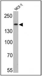
- Western blot analysis of HER-3 was performed by loading 25 µg of MCF-7 cell lysates onto an SDS polyacrylamide gel. Proteins were transferred to a PVDF membrane and blocked at 4ºC overnight. The membrane was probed with a HER-3 monoclonal antibody (Product # MA5-13008) at a dilution of 1:10 overnight at 4°C, washed in TBST, and probed with an HRP-conjugated secondary antibody for 1 hr at room temperature in the dark. Chemiluminescent detection was performed using Pierce ECL Plus Western Blotting Substrate (Product # 32132). Results show a band at ~185 kDa.

Supportive validation
- Submitted by
- Invitrogen Antibodies (provider)
- Main image
- Experimental details
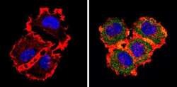
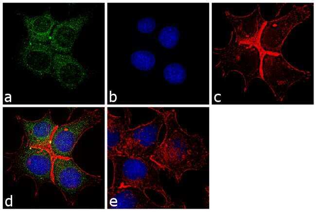
- Immunofluorescent analysis of HER-3 (green) showing staining in the cytoplasm of MCF-7 cells (right) compared to a negative control without primary antibody (left). Formalin-fixed cells were permeabilized with 0.1% Triton X-100 in TBS for 5-10 minutes and blocked with 3% BSA-PBS for 30 minutes at room temperature. Cells were probed with a HER-3 monoclonal antibody (Product # MA5-13008) in 3% BSA-PBS at a dilution of 1:20 and incubated overnight at 4 ºC in a humidified chamber. Cells were washed with PBST and incubated with a DyLight-conjugated secondary antibody in PBS at room temperature in the dark. F-actin (red) was stained with a fluorescent red phalloidin and nuclei (blue) were stained with Hoechst or DAPI. Images were taken at a magnification of 60x.
- Submitted by
- Invitrogen Antibodies (provider)
- Main image
- Experimental details
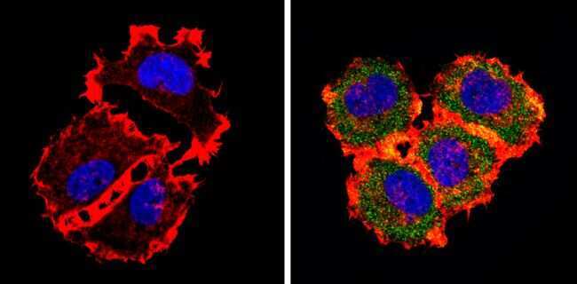
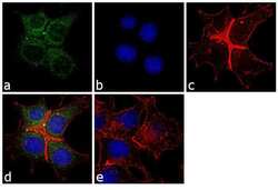
- Immunofluorescence analysis of HER-3 was performed using 70% confluent log phase MCF-7 cells. The cells were fixed with 4% paraformaldehyde for 10 minutes, permeabilized with 0.1% Triton™ X-100 for 10 minutes, and blocked with 1% BSA for 1 hour at room temperature. The cells were labeled with ErbB3 (H3.105.5 (Ab105)) Mouse Monoclonal Antibody (Product # MA5-13008) at 2 µg/mL in 0.1% BSA and incubated for 3 hours at room temperature and then labeled with Goat anti-Mouse IgG (H+L) Superclonal™ Secondary Antibody, Alexa Fluor® 488 conjugate (Product # A28175) at a dilution of 1:2000 for 45 minutes at room temperature (Panel a: green). Nuclei (Panel b: blue) were stained with SlowFade® Gold Antifade Mountant with DAPI (Product # S36938). F-actin (Panel c: red) was stained with Rhodamine Phalloidin (Product # R415, 1:300). Panel d represents the merged image showing cytoplasmic localization. Panel e shows the no primary antibody control. The images were captured at 60X magnification.
Supportive validation
- Submitted by
- Invitrogen Antibodies (provider)
- Main image

- Experimental details
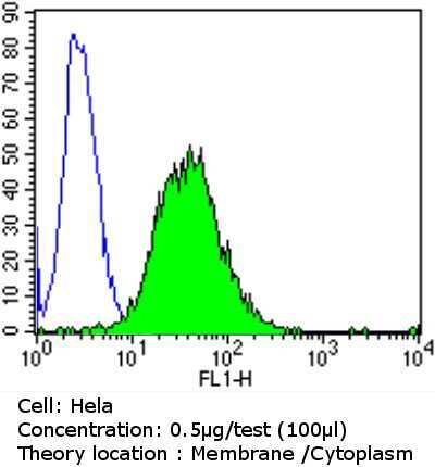
- Flow cytometry analysis of HER-3 in Hela cells compared to an isotype control (blue). Cells were harvested, adjusted to a concentration of 1-5x10^6 cells/mL, fixed with 2% paraformaldehyde and washed with PBS. Cells were blocked with a 2% solution of BSA-PBS for 30 min at room temperature and incubated with a HER-3 monoclonal antibody (Product # MA5-13008) at a dilution of 0.5 µg/test for 60 min at room temperature. Cells were then incubated for 40 min at room temperature in the dark using a Dylight 488-conjugated goat anti-mouse IgG (H+L) secondary antibody and re-suspended in PBS for FACS analysis.
- Submitted by
- Invitrogen Antibodies (provider)
- Main image
- Experimental details
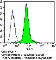
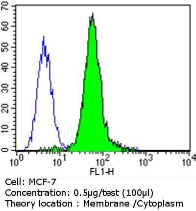
- Flow cytometry analysis of HER-3 in MCF-7 cells compared to an isotype control (blue). Cells were harvested, adjusted to a concentration of 1-5x10^6 cells/mL, fixed with 2% paraformaldehyde and washed with PBS. Cells were blocked with a 2% solution of BSA-PBS for 30 min at room temperature and incubated with a HER-3 monoclonal antibody (Product # MA5-13008) at a dilution of 0.5 µg/test for 60 min at room temperature. Cells were then incubated for 40 min at room temperature in the dark using a Dylight 488-conjugated goat anti-mouse IgG (H+L) secondary antibody and re-suspended in PBS for FACS analysis.
- Submitted by
- Invitrogen Antibodies (provider)
- Main image
- Experimental details
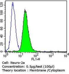
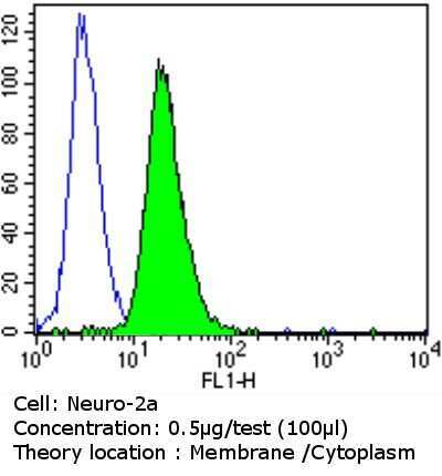
- Flow cytometry analysis of HER-3 in Neuro-2a cells compared to an isotype control (blue). Cells were harvested, adjusted to a concentration of 1-5x10^6 cells/mL, fixed with 2% paraformaldehyde and washed with PBS. Cells were blocked with a 2% solution of BSA-PBS for 30 min at room temperature and incubated with a HER-3 monoclonal antibody (Product # MA5-13008) at a dilution of 0.5 µg/test for 60 min at room temperature. Cells were then incubated for 40 min at room temperature in the dark using a Dylight 488-conjugated goat anti-mouse IgG (H+L) secondary antibody and re-suspended in PBS for FACS analysis.
Supportive validation
- Submitted by
- Invitrogen Antibodies (provider)
- Main image
- Experimental details
- NULL
- Submitted by
- Invitrogen Antibodies (provider)
- Main image

- Experimental details
- NULL