Explore
 Validate
 Learn
Learn Western blot
Antibody data
- Antibody Data
- Antigen structure
- References [27]
- Comments [0]
- Validations
- Western blot [2]
- Other assay [2]
Submit
Validation data
Reference
Comment
Report error
- Product number
- MA5-12675 - Provider product page

- Provider
- Invitrogen Antibodies
- Product name
- ErbB3 Monoclonal Antibody (2F12)
- Antibody type
- Monoclonal
- Antigen
- Recombinant full-length protein
- Description
- MA5-12675 targets HER-3 in WB applications and shows reactivity with Bovine, Human, mouse, and Rat samples. The MA5-12675 immunogen is recombinant rat c-erbB-3/HER-3 oncoprotein.
- Reactivity
- Human, Mouse, Rat, Bovine
- Host
- Mouse
- Isotype
- IgG
- Antibody clone number
- 2F12
- Vial size
- 500 μL
- Concentration
- 0.2 mg/mL
- Storage
- 4°C
Submitted references A bivalent, bispecific Dab-Fc antibody molecule for dual targeting of HER2 and HER3.
Inhibition of Tumor Cell Growth and Cancer Stem Cell Expansion by a Bispecific Antibody Targeting EGFR and HER3.
Additive impact of HER2-/PTK6-RNAi on interactions with HER3 or IGF-1R leads to reduced breast cancer progression in vivo.
Ligand-associated ERBB2/3 activation confers acquired resistance to FGFR inhibition in FGFR3-dependent cancer cells.
EGFR, HER2 and HER3 dimerization patterns guide targeted inhibition in two histotypes of esophageal cancer.
HER2/HER3 heterodimers and p21 expression are capable of predicting adjuvant trastuzumab response in HER2+ breast cancer.
T47D breast cancer cells switch from ER/HER to HER/c-Src signaling upon acquiring resistance to the antiestrogen fulvestrant.
Epithelial-mesenchymal transition markers and HER3 expression are predictors of elisidepsin treatment response in breast and pancreatic cancer cell lines.
MiR-221/-222 differentiate prognostic groups in advanced breast cancers and influence cell invasion.
Spontaneous and pronase-induced HER2 truncation increases the trastuzumab binding capacity of breast cancer tissues and cell lines.
Strong EGFR signaling in cell line models of ERBB2-amplified breast cancer attenuates response towards ERBB2-targeting drugs.
In situ detection of HER2:HER2 and HER2:HER3 protein-protein interactions demonstrates prognostic significance in early breast cancer.
HER-2 signaling, acquisition of growth factor independence, and regulation of biological networks associated with cell transformation.
An activated ErbB3/NRG1 autocrine loop supports in vivo proliferation in ovarian cancer cells.
EGFR over-expression and activation in high HER2, ER negative breast cancer cell line induces trastuzumab resistance.
All EGF(ErbB) receptors have preformed homo- and heterodimeric structures in living cells.
HER kinase activation confers resistance to MET tyrosine kinase inhibition in MET oncogene-addicted gastric cancer cells.
ErbB4 regulates fetal surfactant phospholipid synthesis in primary fetal rat type II cells.
Growth stimulation of non-small cell lung cancer cell lines by antibody against epidermal growth factor receptor promoting formation of ErbB2/ErbB3 heterodimers.
ErbB receptor dimerization, localization, and co-localization in mouse lung type II epithelial cells.
Therapeutic targeting of multiple signaling pathways in malignant pleural mesothelioma.
The neuregulin GGF2 attenuates free radical release from activated microglial cells.
Roles of mitogen-activated protein kinase and phosphoinositide 3'-kinase in ErbB2/ErbB3 coreceptor-mediated heregulin signaling.
The tyrosine kinase inhibitor ZD1839 ("Iressa") inhibits HER2-driven signaling and suppresses the growth of HER2-overexpressing tumor cells.
Reciprocal signaling between spiral ganglion neurons and Schwann cells involves neuregulin and neurotrophins.
Heregulin-dependent activation of phosphoinositide 3-kinase and Akt via the ErbB2/ErbB3 co-receptor.
Heregulin-stimulated signaling in rat pheochromocytoma cells. Evidence for ErbB3 interactions with Neu/ErbB2 and p85.
Rau A, Kocher K, Rommel M, Kühl L, Albrecht M, Gotthard H, Aschmoneit N, Noll B, Olayioye MA, Kontermann RE, Seifert O
mAbs 2021 Jan-Dec;13(1):1902034
mAbs 2021 Jan-Dec;13(1):1902034
Inhibition of Tumor Cell Growth and Cancer Stem Cell Expansion by a Bispecific Antibody Targeting EGFR and HER3.
Rau A, Lieb WS, Seifert O, Honer J, Birnstock D, Richter F, Aschmoneit N, Olayioye MA, Kontermann RE
Molecular cancer therapeutics 2020 Jul;19(7):1474-1485
Molecular cancer therapeutics 2020 Jul;19(7):1474-1485
Additive impact of HER2-/PTK6-RNAi on interactions with HER3 or IGF-1R leads to reduced breast cancer progression in vivo.
Falkenberg N, Anastasov N, Höfig I, Bashkueva K, Lindner K, Höfler H, Rosemann M, Aubele M
Molecular oncology 2015 Jan;9(1):282-94
Molecular oncology 2015 Jan;9(1):282-94
Ligand-associated ERBB2/3 activation confers acquired resistance to FGFR inhibition in FGFR3-dependent cancer cells.
Wang J, Mikse O, Liao RG, Li Y, Tan L, Janne PA, Gray NS, Wong KK, Hammerman PS
Oncogene 2015 Apr 23;34(17):2167-77
Oncogene 2015 Apr 23;34(17):2167-77
EGFR, HER2 and HER3 dimerization patterns guide targeted inhibition in two histotypes of esophageal cancer.
Fichter CD, Timme S, Braun JA, Gudernatsch V, Schöpflin A, Bogatyreva L, Geddert H, Faller G, Klimstra D, Tang L, Hauschke D, Werner M, Lassmann S
International journal of cancer 2014 Oct 1;135(7):1517-30
International journal of cancer 2014 Oct 1;135(7):1517-30
HER2/HER3 heterodimers and p21 expression are capable of predicting adjuvant trastuzumab response in HER2+ breast cancer.
Green AR, Barros FF, Abdel-Fatah TM, Moseley P, Nolan CC, Durham AC, Rakha EA, Chan S, Ellis IO
Breast cancer research and treatment 2014 May;145(1):33-44
Breast cancer research and treatment 2014 May;145(1):33-44
T47D breast cancer cells switch from ER/HER to HER/c-Src signaling upon acquiring resistance to the antiestrogen fulvestrant.
Kirkegaard T, Hansen SK, Larsen SL, Reiter BE, Sørensen BS, Lykkesfeldt AE
Cancer letters 2014 Mar 1;344(1):90-100
Cancer letters 2014 Mar 1;344(1):90-100
Epithelial-mesenchymal transition markers and HER3 expression are predictors of elisidepsin treatment response in breast and pancreatic cancer cell lines.
Teixidó C, Marés R, Aracil M, Ramón y Cajal S, Hernández-Losa J
PloS one 2013;8(1):e53645
PloS one 2013;8(1):e53645
MiR-221/-222 differentiate prognostic groups in advanced breast cancers and influence cell invasion.
Falkenberg N, Anastasov N, Rappl K, Braselmann H, Auer G, Walch A, Huber M, Höfig I, Schmitt M, Höfler H, Atkinson MJ, Aubele M
British journal of cancer 2013 Nov 12;109(10):2714-23
British journal of cancer 2013 Nov 12;109(10):2714-23
Spontaneous and pronase-induced HER2 truncation increases the trastuzumab binding capacity of breast cancer tissues and cell lines.
Recupero D, Daniele L, Marchiò C, Molinaro L, Castellano I, Cassoni P, Righi A, Montemurro F, Sismondi P, Biglia N, Viale G, Risio M, Sapino A
The Journal of pathology 2013 Feb;229(3):390-9
The Journal of pathology 2013 Feb;229(3):390-9
Strong EGFR signaling in cell line models of ERBB2-amplified breast cancer attenuates response towards ERBB2-targeting drugs.
Henjes F, Bender C, von der Heyde S, Braun L, Mannsperger HA, Schmidt C, Wiemann S, Hasmann M, Aulmann S, Beissbarth T, Korf U
Oncogenesis 2012 Jul 2;1(7):e16
Oncogenesis 2012 Jul 2;1(7):e16
In situ detection of HER2:HER2 and HER2:HER3 protein-protein interactions demonstrates prognostic significance in early breast cancer.
Spears M, Taylor KJ, Munro AF, Cunningham CA, Mallon EA, Twelves CJ, Cameron DA, Thomas J, Bartlett JM
Breast cancer research and treatment 2012 Apr;132(2):463-70
Breast cancer research and treatment 2012 Apr;132(2):463-70
HER-2 signaling, acquisition of growth factor independence, and regulation of biological networks associated with cell transformation.
Bollig-Fischer A, Dziubinski M, Boyer A, Haddad R, Giroux CN, Ethier SP
Cancer research 2010 Oct 15;70(20):7862-73
Cancer research 2010 Oct 15;70(20):7862-73
An activated ErbB3/NRG1 autocrine loop supports in vivo proliferation in ovarian cancer cells.
Sheng Q, Liu X, Fleming E, Yuan K, Piao H, Chen J, Moustafa Z, Thomas RK, Greulich H, Schinzel A, Zaghlul S, Batt D, Ettenberg S, Meyerson M, Schoeberl B, Kung AL, Hahn WC, Drapkin R, Livingston DM, Liu JF
Cancer cell 2010 Mar 16;17(3):298-310
Cancer cell 2010 Mar 16;17(3):298-310
EGFR over-expression and activation in high HER2, ER negative breast cancer cell line induces trastuzumab resistance.
Dua R, Zhang J, Nhonthachit P, Penuel E, Petropoulos C, Parry G
Breast cancer research and treatment 2010 Aug;122(3):685-97
Breast cancer research and treatment 2010 Aug;122(3):685-97
All EGF(ErbB) receptors have preformed homo- and heterodimeric structures in living cells.
Tao RH, Maruyama IN
Journal of cell science 2008 Oct 1;121(Pt 19):3207-17
Journal of cell science 2008 Oct 1;121(Pt 19):3207-17
HER kinase activation confers resistance to MET tyrosine kinase inhibition in MET oncogene-addicted gastric cancer cells.
Bachleitner-Hofmann T, Sun MY, Chen CT, Tang L, Song L, Zeng Z, Shah M, Christensen JG, Rosen N, Solit DB, Weiser MR
Molecular cancer therapeutics 2008 Nov;7(11):3499-508
Molecular cancer therapeutics 2008 Nov;7(11):3499-508
ErbB4 regulates fetal surfactant phospholipid synthesis in primary fetal rat type II cells.
Zscheppang K, Liu W, Volpe MV, Nielsen HC, Dammann CE
American journal of physiology. Lung cellular and molecular physiology 2007 Aug;293(2):L429-35
American journal of physiology. Lung cellular and molecular physiology 2007 Aug;293(2):L429-35
Growth stimulation of non-small cell lung cancer cell lines by antibody against epidermal growth factor receptor promoting formation of ErbB2/ErbB3 heterodimers.
Maegawa M, Takeuchi K, Funakoshi E, Kawasaki K, Nishio K, Shimizu N, Ito F
Molecular cancer research : MCR 2007 Apr;5(4):393-401
Molecular cancer research : MCR 2007 Apr;5(4):393-401
ErbB receptor dimerization, localization, and co-localization in mouse lung type II epithelial cells.
Zscheppang K, Korenbaum E, Bueter W, Ramadurai SM, Nielsen HC, Dammann CE
Pediatric pulmonology 2006 Dec;41(12):1205-12
Pediatric pulmonology 2006 Dec;41(12):1205-12
Therapeutic targeting of multiple signaling pathways in malignant pleural mesothelioma.
Mukohara T, Civiello G, Johnson BE, Janne PA
Oncology 2005;68(4-6):500-10
Oncology 2005;68(4-6):500-10
The neuregulin GGF2 attenuates free radical release from activated microglial cells.
Dimayuga FO, Ding Q, Keller JN, Marchionni MA, Seroogy KB, Bruce-Keller AJ
Journal of neuroimmunology 2003 Mar;136(1-2):67-74
Journal of neuroimmunology 2003 Mar;136(1-2):67-74
Roles of mitogen-activated protein kinase and phosphoinositide 3'-kinase in ErbB2/ErbB3 coreceptor-mediated heregulin signaling.
Vijapurkar U, Kim MS, Koland JG
Experimental cell research 2003 Apr 1;284(2):291-302
Experimental cell research 2003 Apr 1;284(2):291-302
The tyrosine kinase inhibitor ZD1839 ("Iressa") inhibits HER2-driven signaling and suppresses the growth of HER2-overexpressing tumor cells.
Moasser MM, Basso A, Averbuch SD, Rosen N
Cancer research 2001 Oct 1;61(19):7184-8
Cancer research 2001 Oct 1;61(19):7184-8
Reciprocal signaling between spiral ganglion neurons and Schwann cells involves neuregulin and neurotrophins.
Hansen MR, Vijapurkar U, Koland JG, Green SH
Hearing research 2001 Nov;161(1-2):87-98
Hearing research 2001 Nov;161(1-2):87-98
Heregulin-dependent activation of phosphoinositide 3-kinase and Akt via the ErbB2/ErbB3 co-receptor.
Hellyer NJ, Kim MS, Koland JG
The Journal of biological chemistry 2001 Nov 9;276(45):42153-61
The Journal of biological chemistry 2001 Nov 9;276(45):42153-61
Heregulin-stimulated signaling in rat pheochromocytoma cells. Evidence for ErbB3 interactions with Neu/ErbB2 and p85.
Gamett DC, Greene T, Wagreich AR, Kim HH, Koland JG, Cerione RA
The Journal of biological chemistry 1995 Aug 11;270(32):19022-7
The Journal of biological chemistry 1995 Aug 11;270(32):19022-7
No comments: Submit comment
Supportive validation
- Submitted by
- Invitrogen Antibodies (provider)
- Main image
- Experimental details
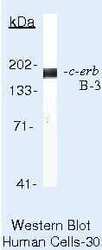
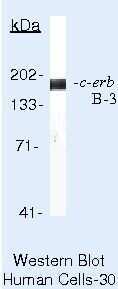
- Western blot of HER-3 using HER-3 Monoclonal Antibody (Product # MA5-12675) on T47D Cells.
- Submitted by
- Invitrogen Antibodies (provider)
- Main image
- Experimental details
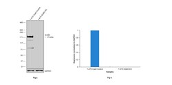
- Knockout of ErbB3 was achieved by CRISPR-Cas9 genome editing using LentiArray™ Lentiviral sgRNA (Product # A32042, Assay ID CRISPR764358_LV) and LentiArray Cas9 Lentivirus (Product # A32064). Western blot analysis of ErbB3 was performed by loading 30 µg of T-47D Cas9 (Lane 1) andT-47D ErbB3 KO (Lane 2) membrane enriched extracts. The samples were electrophoresed using NuPAGE™ 3-8% Tris-Acetate Protein Gel (Product # EA0378BOX). Resolved proteins were then transferred onto a nitrocellulose membrane (Product # IB23001) by iBlot® 2 Dry Blotting System (Product # IB21001). The blot was probed with Anti-ErbB3 Monoclonal Antibody (2F12) (Product # MA5-12675, 4 µg/mL dilution) and Goat anti-Mouse IgG (H+L) Superclonal™ Recombinant Secondary Antibody, HRP (Product # A28177, 1:5000 dilution) using the iBright FL 1000 (Product # A32752). Chemiluminescent detection was performed using SuperSignal™ West Dura Extended Duration Substrate (Product # 34076). Loss of signal upon CRISPR mediated knockout (KO) using the LentiArray™ CRISPR product line confirms that antibody is specific to ErbB3. Uncharacterized bands were observed at ~80 kDa and ~150 kDa in T-47D Cas9 sample.

Supportive validation
- Submitted by
- Invitrogen Antibodies (provider)
- Main image
- Experimental details
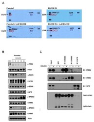
- Fig. 2 Activation of ERBB2 and ERBB3 in BGJ398 RS cells (A) A phospho-RTK array reveals that BGJ398 RS cells have increased phosphorylation of ERBB2 and ERBB3 in the presence of BGJ398. The cell lysates were hybridized to a phospho-RTK membrane, on which each RTK is spotted in duplicate. Hybridization signals at the corners serve as controls. (B) Immunoblot analysis of protein phosphorylation in extracts from parental cells that were treated for the indicated time points with 1muM BGJ398. (C) ERBB2 and ERBB3 dimerization measured by co-immunoprecipitation. The parental cells were treated with 1muM BGJ398 for 24h and prior to lysis. The experiments were performed at least twice.

- Submitted by
- Invitrogen Antibodies (provider)
- Main image
- Experimental details
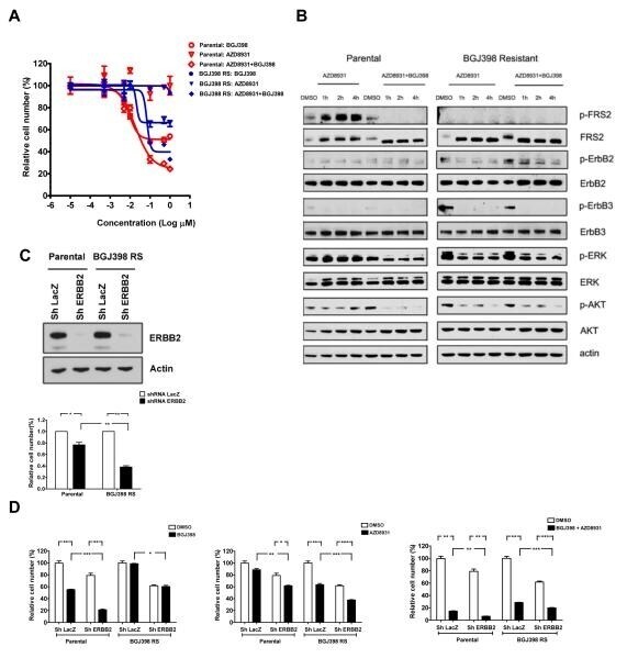
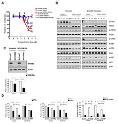
- Fig. 3 BGJ398 RS cell lines are selectively sensitive to tyrosine kinase inhibitors and to shRNA-mediated depletion of ERBB2 or ERBB3 A) BGJ398 RS cells are sensitive to the ERBB family inhibitor. Parental and BGJ398 RS cells were exposed to BGJ398 or AZD8931 alone or in combination, and cell proliferation was measured 4 days after treatment using Cell-Titer-GLO. (B) Immunoblot analysis of protein phosphorylation in extracts from parental and BGJ398 RS cells that were treated for indicated time point with 1muM BGJ398, 1muM AZD8931 or a combination of both drugs. (C) Upper: Immunoblots demonstrate down-regulation of ERBB2 by the specific shRNA. A lacZ control or ERBB2 specific shRNAs were introduced into parental or BGJ398 RS cells. Cells underwent puromycin selection and extracts were immunoblotted with the indicated antibodies. Lower: BGJ398 RS cells are sensitive to knock-down of ERBB2 expression. The number of cells were counted 72 hours after seeding, and the viability of knockdown samples (in triplicate) is presented relative to LacZ control sample. (D) Knockdown of ERBB2 also sensitizes the BGJ398 RS cells to AZD8931. Parental and BGJ398 RS cells were infected with indicated shRNA and the proliferation is shown for indicated cells treated for 96 hours with 1muM BGJ398, 1muM AZD8931 or a combination of both drugs. Proliferation is shown relative to untreated cells at the same time point. The experiments were repeated twice. *, P