Explore
 Validate
 Learn
LearnPB9917
antibody from Boster Biological Technology
Targeting: BCL2L1
Bcl-X, bcl-xL, bcl-xS, BCL2L, BCLX, PPP1R52
 Western blot
 Immunocytochemistry
Antibody data
- Antibody Data
- Antigen structure
- References [5]
- Comments [0]
- Validations
- Western blot [1]
Submit
Validation data
Reference
Comment
Report error
- Product number
- PB9917 - Provider product page

- Provider
- Boster Biological Technology
- Product name
- Anti-Bcl-X/BCL2L1 Antibody Picoband™
- Antibody type
- Polyclonal
- Description
- Polyclonal antibody for BCL XL/BCL2L1 detection. Host: Rabbit.Size: 100μg/vial. Tested applications: WB. Reactive species: Human. BCL XL/BCL2L1 information: Molecular Weight: 26049 MW; Subcellular Localization: Isoform Bcl-X(L): Mitochondrion inner membrane . Mitochondrion outer membrane . Mitochondrion matrix . Cytoplasmic vesicle, secretory vesicle, synaptic vesicle membrane . Cytoplasm, cytosol . Cytoplasm, cytoskeleton, microtubule organizing center, centrosome. Nucleus membrane ; Single-pass membrane protein ; Cytoplasmic side . After neuronal stimulation, translocates from cytosol to synaptic vesicle and mitochondrion membrane in a calmodulin-dependent manner (By similarity). Localizes to the centrosome when phosphorylated at Ser-49; Tissue Specificity: Bcl-X(S) is expressed at high levels in cells that undergo a high rate of turnover, such as developing lymphocytes. In contrast, Bcl-X(L) is found in tissues containing long-lived postmitotic cells, such as adult brain.
- Reactivity
- Human
- Host
- Rabbit
- Vial size
- 100μg/vial
- Concentration
- Add 0.2ml of distilled water will yield a concentration of 500ug/ml.
- Storage
- At -20°C for one year. After reconstitution, at 4°C for one month. It can also be aliquoted and stored frozen at -20°C for a longer time. Avoid repeated freezing and thawing.
- Handling
- Add 0.2ml of distilled water will yield a concentration of 500ug/ml.
Submitted references The Inhibition of Gastric Cancer Cells' Progression by 23,24-Dihydrocucurbitacin E through Disruption of the Ras/Raf/ERK/MMP9 Signaling Pathway.
Active targeting co-delivery system based on hollow mesoporous silica nanoparticles for antitumor therapy in ovarian cancer stem-like cells.
The role of cofilin-l in vulvar squamous cell carcinoma: A marker of carcinogenesis, progression and targeted therapy.
Aspirin overcomes Navitoclax-resistance in hepatocellular carcinoma cells through suppression of Mcl-1.
ACTX-8, a cytotoxic L-amino acid oxidase isolated from Agkistrodon acutus snake venom, induces apoptosis in Hela cervical cancer cells.
Liu H, Wang H, Dong A, Huo X, Wang H, Wang J, Si J
Molecules (Basel, Switzerland) 2022 Apr 22;27(9)
Molecules (Basel, Switzerland) 2022 Apr 22;27(9)
Active targeting co-delivery system based on hollow mesoporous silica nanoparticles for antitumor therapy in ovarian cancer stem-like cells.
Guo X, Guo N, Zhao J, Cai Y
Oncology reports 2017 Sep;38(3):1442-1450
Oncology reports 2017 Sep;38(3):1442-1450
The role of cofilin-l in vulvar squamous cell carcinoma: A marker of carcinogenesis, progression and targeted therapy.
Wu Q, Jiang Y, Cui S, Wang Y, Wu X
Oncology reports 2016 May;35(5):2743-54
Oncology reports 2016 May;35(5):2743-54
Aspirin overcomes Navitoclax-resistance in hepatocellular carcinoma cells through suppression of Mcl-1.
Li G, Zhang S, Fang H, Yan B, Zhao Y, Feng L, Ma X, Ye X
Biochemical and biophysical research communications 2013 May 17;434(4):809-14
Biochemical and biophysical research communications 2013 May 17;434(4):809-14
ACTX-8, a cytotoxic L-amino acid oxidase isolated from Agkistrodon acutus snake venom, induces apoptosis in Hela cervical cancer cells.
Zhang L, Wei LJ
Life sciences 2007 Mar 6;80(13):1189-97
Life sciences 2007 Mar 6;80(13):1189-97
No comments: Submit comment
Supportive validation
- Submitted by
- Boster Biological Technology (provider)
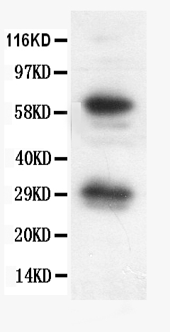
- Main image
- Experimental details
- Western blot analysis of Bcl-X using anti- Bcl-X antibody (PB9917). Electrophoresis was performed on a 5-20% SDS-PAGE gel at 70V (Stacking gel) / 90V (Resolving gel) for 2-3 hours. The sample well of each lane was loaded with 50ug of sample under reducing conditions. Lane 1: SW620 whole cell lysates. After Electrophoresis, proteins were transferred to a Nitrocellulose membrane at 150mA for 50-90 minutes. Blocked the membrane with 5% Non-fat Milk/ TBS for 1.5 hour at RT. The membrane was incubated with rabbit anti- Bcl-X antigen affinity purified polyclonal antibody (Catalog # PB9917) at 0.5 μg/mL overnight at 4°C, then washed with TBS-0.1%Tween 3 times with 5 minutes each and probed with a goat anti-rabbit IgG-HRP secondary antibody at a dilution of 1:10000 for 1.5 hour at RT. The signal is developed using an Enhanced Chemiluminescent detection (ECL) kit (Catalog # EK1002) with Tanon 5200 system. A specific band was detected for Bcl-X at approximately 29 KD, 60KD. The expected band size for Bcl-X is at 26KD.
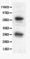
- Additional image