 Explore
 Validate
 Learn
Learn14-6994-81
antibody from Invitrogen Antibodies
Targeting: BCL2L1
Bcl-X, bcl-xL, bcl-xS, BCL2L, BCLX, PPP1R52
 Western blot
Antibody data
- Antibody Data
- Antigen structure
- References [5]
- Comments [0]
- Validations
- Western blot [2]
- Immunocytochemistry [1]
- Other assay [1]
Submit
Validation data
Reference
Comment
Report error
- Product number
- 14-6994-81 - Provider product page

- Provider
- Invitrogen Antibodies
- Product name
- Bcl-X Monoclonal Antibody (2H12), eBioscience™
- Antibody type
- Monoclonal
- Antigen
- Other
- Description
- Description: The 2H12 antibody reacts with human, mouse and rat Bcl-X. Bcl-X functions as a central repressor of several apoptotic signals.This antibody reacts with both Bcl-XS (short) and Bcl-XL (long) proteins.
- Antibody clone number
- 2H12
- Concentration
- 0.5 mg/mL
Submitted references Anticancer Activity of Continentalic Acid in B-Cell Lymphoma.
Heat shock protein 90 maintains the tumour-like character of rheumatoid synovial cells by stabilizing integrin-linked kinase, extracellular signal-regulated kinase and protein kinase B.
Translocation of p53 to mitochondria is regulated by its lipid binding property to anionic phospholipids and it participates in cell death control.
Translocation of p53 to mitochondria is regulated by its lipid binding property to anionic phospholipids and it participates in cell death control.
Nonionic detergents induce dimerization among members of the Bcl-2 family.
Jeon BE, Kwon CS, Lee JE, Moon K, Cha J, Park I, Koh S, Yoon M, Kim SW, Kim JN
Molecules (Basel, Switzerland) 2021 Nov 12;26(22)
Molecules (Basel, Switzerland) 2021 Nov 12;26(22)
Heat shock protein 90 maintains the tumour-like character of rheumatoid synovial cells by stabilizing integrin-linked kinase, extracellular signal-regulated kinase and protein kinase B.
Hashiramoto A, Murata M, Kawazoe T, Yoshida K, Akiyama C, Shiozawa K, Shiozawa S
Rheumatology (Oxford, England) 2011 May;50(5):852-61
Rheumatology (Oxford, England) 2011 May;50(5):852-61
Translocation of p53 to mitochondria is regulated by its lipid binding property to anionic phospholipids and it participates in cell death control.
Li CH, Cheng YW, Liao PL, Kang JJ
Neoplasia (New York, N.Y.) 2010 Feb;12(2):150-60
Neoplasia (New York, N.Y.) 2010 Feb;12(2):150-60
Translocation of p53 to mitochondria is regulated by its lipid binding property to anionic phospholipids and it participates in cell death control.
Li CH, Cheng YW, Liao PL, Kang JJ
Neoplasia (New York, N.Y.) 2010 Feb;12(2):150-60
Neoplasia (New York, N.Y.) 2010 Feb;12(2):150-60
Nonionic detergents induce dimerization among members of the Bcl-2 family.
Hsu YT, Youle RJ
The Journal of biological chemistry 1997 May 23;272(21):13829-34
The Journal of biological chemistry 1997 May 23;272(21):13829-34
No comments: Submit comment
Supportive validation
- Submitted by
- Invitrogen Antibodies (provider)
- Main image
- Experimental details
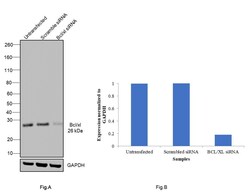
- Knockdown of Bcl-X was achieved by transfecting A549 with Bcl-X specific siRNAs (Silencer® select Product # S1921, S1920). Western blot analysis (Fig. a) was performed using Whole cell extracts from the Bcl-Xl untransfected cells (lane 1), non-targeting scrambled siRNA transfected cells (lane 2) and knockdown cells (lane 3). The blot was probed with Bcl-X Monoclonal Antibody (2H12), eBioscience™ (Product # 14-6994-81, 1:500 dilution) and Goat anti-Mouse IgG (H+L) Superclonal™ Recombinant Secondary Antibody, HRP (Product # A28177, 1:4000 dilution). Densitometric analysis of this western blot is shown in histogram (Fig. b). Decrease in signal upon siRNA mediated knock down confirms that antibody is specific to Bcl-X.

- Submitted by
- Invitrogen Antibodies (provider)
- Main image
- Experimental details
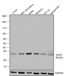
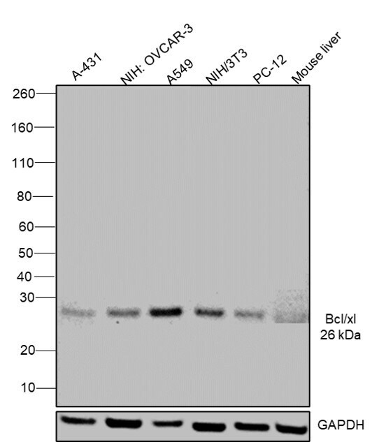
- Western blot was performed using Anti-Bcl-X Monoclonal Antibody (2H12), eBioscience™ (Product # 14-6994-81) and a 26 kDa band corresponding to Bcl-X was observed across cell lines and tissues tested. Whole cell extracts (30 µg lysate) of A-431 (Lane 1), NIH:OVCAR-3 (Lane 2), A549 (Lane 3), NIH/3T3 (Lane 4),PC-12 (Lane 5) and mouse liver(Lane 6) were electrophoresed using NuPAGE™ 4-12% Bis-Tris Protein Gel (Product # NP0321BOX). Resolved proteins were then transferred onto a Nitrocellulose membrane (Product # IB23001) by iBlot® 2 Dry Blotting System (Product # IB21001). The blot was probed with the primary antibody (1 µg/mL concentration) and detected by chemiluminescence with Goat anti-Mouse IgG (H+L) Superclonal™ Recombinant Secondary Antibody, HRP (Product # A28177, 1:4000 dilution) using the iBright FL 1000 (Product # A32752). Chemiluminescent detection was performed using Novex® ECL Chemiluminescent Substrate Reagent Kit (Product # WP20005).
Supportive validation
- Submitted by
- Invitrogen Antibodies (provider)
- Main image

- Experimental details
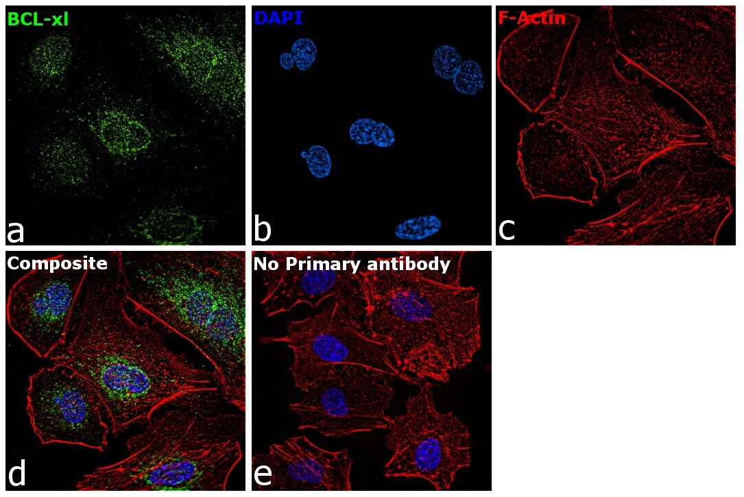
- Immunofluorescence analysis of Bcl-X was performed using 70% confluent log phase A549 cells. The cells were fixed with 4% paraformaldehyde for 10 minutes, permeabilized with 0.1% Triton™ X-100 for 15 minutes, and blocked with 2% BSA for 45 minutes at room temperature. The cells were labeled with Bcl-X Monoclonal Antibody (2H12), eBioscience™ (Product # 14-6994-81) at 5 µg/mL in 0.1% BSA, incubated at 4 degree celsius overnight and then labeled with Goat anti-Mouse IgG (H+L) Superclonal™ Recombinant Secondary Antibody, Alexa Fluor® 488 conjugate (Product # A28175), (1:2000), for 45 minutes at room temperature (Panel a: Green). Nuclei (Panel b:Blue) were stained with ProLong™ Diamond Antifade Mountant with DAPI (Product # P36962). F-actin (Panel c: Red) was stained with Rhodamine Phalloidin (Product # R415, 1:300 dilution). Panel d represents the merged image showing Cytoplasm localization. Panel e represents control cells with no primary antibody to assess background. The images were captured at 60X magnification.
Supportive validation
- Submitted by
- Invitrogen Antibodies (provider)
- Main image
- Experimental details
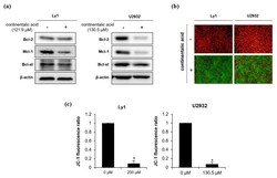
- Figure 6 Continentalic acid reduces expression of anti-apoptotic Bcl-2 family members and dissociates mitochondrial membrane potential (MMP). ( a ) Continentalic acid was added in Ly1 and U2932 cells, and the expression of anti-apoptotic Bcl-2 family members Bcl-2, Mcl-1, and Bcl-xl was detected by Western blot 24 h later. beta-actin served as a control for equal loading. A representative of three independent experiments is shown. ( b ) Ly1 and U2932 were treated with continentalic acid for 24 h and stained with JC-1, followed by monitoring of MMP by fluorescence microscopy. Red (J-aggregates) and green (J-monomers) colors represent intact and disrupted MMP, respectively. ( c ) MMP was calculated using flow cytometry to monitor the JC-1 fluorescence ratio of J-aggregates (red) and J-monomers (green). Statistical significance was calculated using the two-tailed Mann-Whitney test (* p < 0.05).
