Explore
 Validate
 Learn
LearnNB100-56105
antibody from Novus Biologicals
Targeting: BCL2L1
Bcl-X, bcl-xL, bcl-xS, BCL2L, BCLX, PPP1R52
 Western blot
 Immunohistochemistry
Antibody data
- Antibody Data
- Antigen structure
- References [1]
- Comments [0]
- Validations
- Immunohistochemistry [2]
Submit
Validation data
Reference
Comment
Report error
- Product number
- NB100-56105 - Provider product page

- Provider
- Novus Biologicals
- Proper citation
- Novus Cat#NB100-56105, RRID:AB_837711
- Product name
- Rabbit Polyclonal Bcl-xL Antibody
- Antibody type
- Polyclonal
Submitted references Early processing of Bid and caspase-6, -8, -10, -14 in the canine brain during cardiac arrest and resuscitation.
Krajewska M, Rosenthal RE, Mikolajczyk J, Stennicke HR, Wiesenthal T, Mai J, Naito M, Salvesen GS, Reed JC, Fiskum G, Krajewski S
Experimental neurology 2004 Oct;189(2):261-79
Experimental neurology 2004 Oct;189(2):261-79
No comments: Submit comment
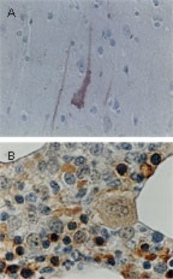
Supportive validation
- Submitted by
- Novus Biologicals (provider)
- Main image
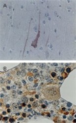
- Experimental details
- Immunohistochemistry-Paraffin: Bcl-xL Antibody [NB100-56105] - FFPE human tissue sections stained for Bcl-X expression using this antibody at 1:2000. (A) Cortex from the brain of an Alzheimer's disease patient. The degenerating neurons are positive for BclX expression, the intact neurons are not. (B) Normal bone marrow. The cells positive for Bcl-X expression appear to be of erythropoetic lineage. Hematoxylin-eosin counterstain.
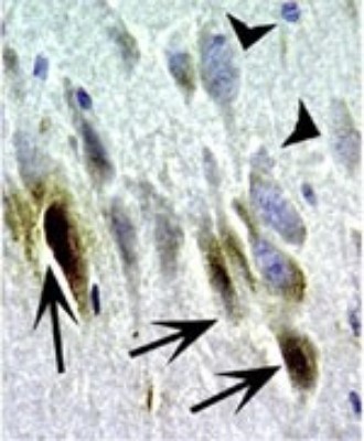
- Submitted by
- Novus Biologicals (provider)
- Main image
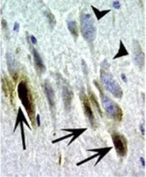
- Experimental details
- Immunohistochemistry-Paraffin: Bcl-xL Antibody [NB100-56105] - FFPE section of dog ischemic brain cortex stained for Bcl-X expression using this antibody at 1:2000. At 2 hr post ischemia, Bcl-X staining was seen in the dying neurons that had morphological features of apoptosis (arrows). In contrast, mophologically normal appearing neurons lacked Bcl-X staining (arrowheads). Hematoxylin-eosin counterstain.