Explore
 Validate
 Learn
LearnHPA002199
antibody from Atlas Antibodies
Targeting: ABCB1
ABC20, CD243, CLCS, GP170, MDR1, P-gp, PGY1
 Immunocytochemistry
Antibody data
- Antibody Data
- Antigen structure
- References [5]
- Comments [0]
- Validations
- Immunocytochemistry [1]
- Immunohistochemistry [1]
Submit
Validation data
Reference
Comment
Report error
- Product number
- HPA002199 - Provider product page

- Provider
- Atlas Antibodies
- Proper citation
- Atlas Antibodies Cat#HPA002199, RRID:AB_1844428
- Product name
- Anti-ABCB1
- Antibody type
- Polyclonal
- Description
- Polyclonal Antibody against Human ABCB1, Gene description: ATP-binding cassette, sub-family B (MDR/TAP), member 1, Alternative Gene Names: ABC20, CD243, CLCS, GP170, MDR1, P-gp, PGY1, Validated applications: ICC, IHC, Uniprot ID: P08183, Storage: Store at +4°C for short term storage. Long time storage is recommended at -20°C.
- Reactivity
- Human
- Host
- Rabbit
- Conjugate
- Unconjugated
- Isotype
- IgG
- Vial size
- 100 µl
- Concentration
- 0.1 mg/ml
- Storage
- Store at +4°C for short term storage. Long time storage is recommended at -20°C.
- Handling
- The antibody solution should be gently mixed before use.
Submitted references Metabolic reprogramming of poly(morpho)nuclear giant cells determines glioblastoma recovery from doxorubicin-induced stress
Key molecular alterations in endothelial cells in human glioblastoma uncovered through single-cell RNA sequencing
Expression of MATE1, P-gp, OCTN1 and OCTN2, in epithelial and immune cells in the lung of COPD and healthy individuals.
ABC-Transporter Expression Does Not Correlate with Response to Irinotecan in Patients with Metastatic Colorectal Cancer.
Vascular and extravascular distribution of the ATP-binding cassette transporters ABCB1 and ABCC1 in aged human brain and pituitary
Pudełek M, Ryszawy D, Piwowarczyk K, Lasota S, Madeja Z, Kędracka-Krok S, Czyż J
Journal of Translational Medicine 2024;22(1)
Journal of Translational Medicine 2024;22(1)
Key molecular alterations in endothelial cells in human glioblastoma uncovered through single-cell RNA sequencing
Xie Y, He L, Lugano R, Zhang Y, Cao H, He Q, Chao M, Liu B, Cao Q, Wang J, Jiao Y, Hu Y, Han L, Zhang Y, Huang H, Uhrbom L, Betsholtz C, Wang L, Dimberg A, Zhang L
JCI Insight 2021
JCI Insight 2021
Expression of MATE1, P-gp, OCTN1 and OCTN2, in epithelial and immune cells in the lung of COPD and healthy individuals.
Berg T, Hegelund-Myrbäck T, Öckinger J, Zhou XH, Brännström M, Hagemann-Jensen M, Werkström V, Seidegård J, Grunewald J, Nord M, Gustavsson L
Respiratory research 2018 Apr 20;19(1):68
Respiratory research 2018 Apr 20;19(1):68
ABC-Transporter Expression Does Not Correlate with Response to Irinotecan in Patients with Metastatic Colorectal Cancer.
Trumpi K, Emmink BL, Prins AM, van Oijen MG, van Diest PJ, Punt CJ, Koopman M, Kranenburg O, Rinkes IH
Journal of Cancer 2015;6(11):1079-86
Journal of Cancer 2015;6(11):1079-86
Vascular and extravascular distribution of the ATP-binding cassette transporters ABCB1 and ABCC1 in aged human brain and pituitary
Bernstein H, Hölzl G, Dobrowolny H, Hildebrandt J, Trübner K, Krohn M, Bogerts B, Pahnke J
Mechanisms of Ageing and Development 2014;141-142
Mechanisms of Ageing and Development 2014;141-142
No comments: Submit comment
Supportive validation
- Submitted by
- Atlas Antibodies (provider)
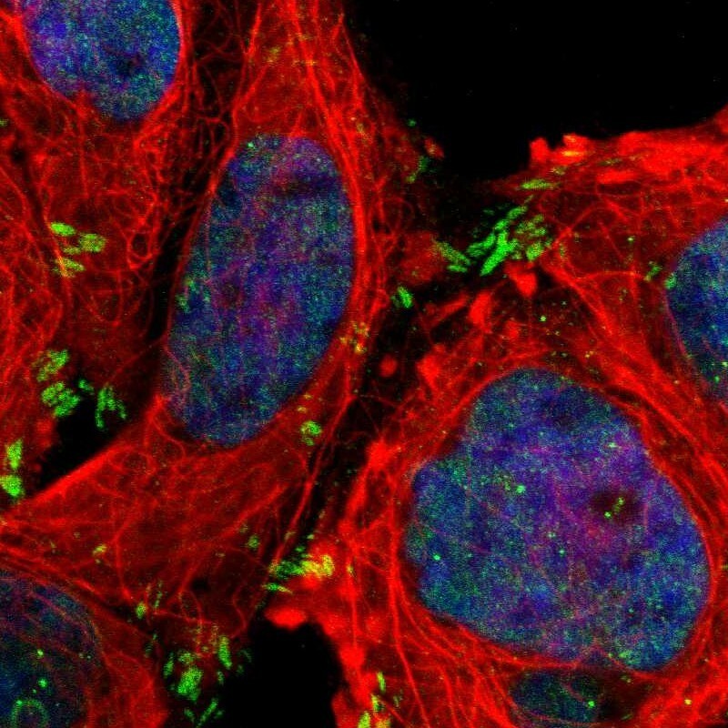
- Main image
- Experimental details
- Immunofluorescent staining of human cell line A-431 shows localization to nucleoplasm & focal adhesion sites.
- Sample type
- Human
Supportive validation
- Submitted by
- Atlas Antibodies (provider)
- Enhanced method
- Orthogonal validation
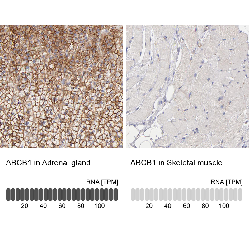
- Main image
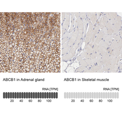
- Experimental details
- Immunohistochemistry analysis in human adrenal gland and skeletal muscle tissues using HPA002199 antibody. Corresponding ABCB1 RNA-seq data are presented for the same tissues.
- Sample type
- Human
- Protocol
- Protocol