Explore
 Validate
 Learn
Learn Western blot
 Immunocytochemistry
Antibody data
- Antibody Data
- Antigen structure
- References [0]
- Comments [0]
- Validations
- Western blot [3]
- Immunocytochemistry [1]
- Immunohistochemistry [1]
Submit
Validation data
Reference
Comment
Report error
- Product number
- GTX132433 - Provider product page

- Provider
- GeneTex
- Product name
- Huntingtin antibody
- Antibody type
- Polyclonal
- Reactivity
- Human, Rat
- Host
- Rabbit
No comments: Submit comment
Enhanced validation
Supportive validation
- Submitted by
- GeneTex (provider)
- Enhanced method
- Genetic validation
- Main image
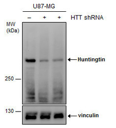
- Experimental details
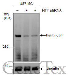
- Non-transfected (¡V) and transfected (+) U87-MG whole cell extracts (30 ?g) were separated by 5% SDS-PAGE, and the membrane was blotted with Huntingtin antibody (GTX132433) diluted at 1:1000.
Supportive validation
- Submitted by
- GeneTex (provider)
- Main image
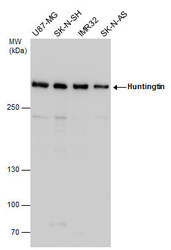
- Experimental details
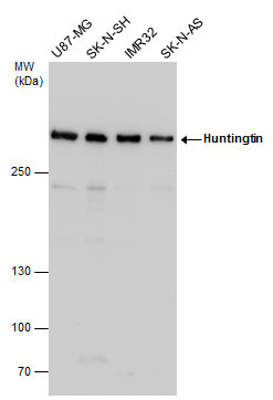
- Various whole cell extracts (30 £gg) were separated by 5% SDS-PAGE, and the membrane was blotted with Huntingtin antibody (GTX132433) diluted at 1:1000.
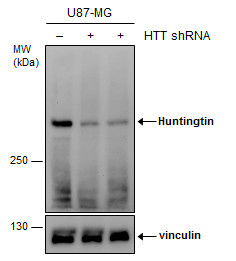
- Submitted by
- GeneTex (provider)
- Main image
- Experimental details
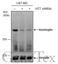
- Non-transfected (¡V) and transfected (+) U87-MG whole cell extracts (30 ?g) were separated by 5% SDS-PAGE, and the membrane was blotted with Huntingtin antibody (GTX132433) diluted at 1:1000.
Supportive validation
- Submitted by
- GeneTex (provider)
- Main image
- Experimental details
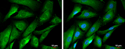
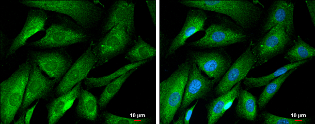
- Huntingtin antibody detects Huntingtin protein at cytoplasm by immunofluorescent analysis.Sample: SK-N-SH cells were fixed in 4% paraformaldehyde at RT for 15 min.Green: Huntingtin protein stained by Huntingtin antibody (GTX132433) diluted at 1:500.Blue: Hoechst 33342 staining.Scale bar = 10 £gm.
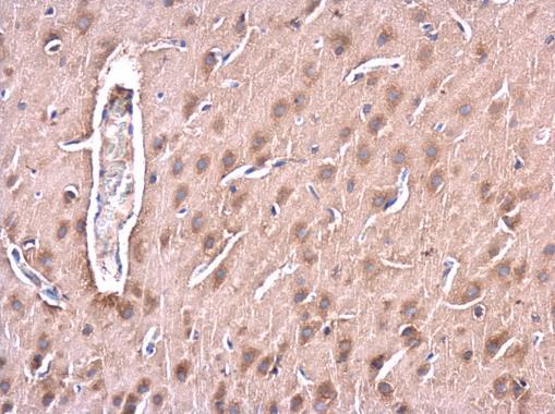
Supportive validation
- Submitted by
- GeneTex (provider)
- Main image
- Experimental details
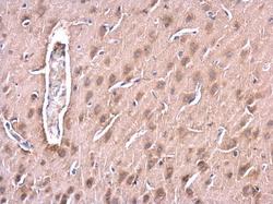
- Huntingtin antibody detects Huntingtin protein at cytoplasm in rat brain by immunohistochemical analysis. Sample: Paraffin-embedded rat brain. Huntingtin antibody (GTX132433) diluted at 1:500.