 Explore
 Validate
 Learn
Learn Western blot
 Immunohistochemistry
Antibody data
- Antibody Data
- Antigen structure
- References [0]
- Comments [0]
- Validations
- Western blot [1]
- Immunocytochemistry [1]
Submit
Validation data
Reference
Comment
Report error
- Product number
- PA1-087 - Provider product page

- Provider
- Invitrogen Antibodies
- Product name
- Phospho-Huntingtin (Ser421) Polyclonal Antibody
- Antibody type
- Polyclonal
- Antigen
- Synthetic peptide
- Description
- PA1-087 has been successfully used in immunofluorescence, immunohistochemistry, and Western blotting applications on human and mouse samples.
- Reactivity
- Human, Mouse
- Host
- Rabbit
- Isotype
- IgG
- Vial size
- 100 µL
- Concentration
- 0.24 mg/mL
- Storage
- -20°C
No comments: Submit comment
Supportive validation
- Submitted by
- Invitrogen Antibodies (provider)
- Main image
- Experimental details
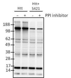
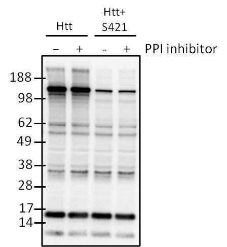
- Western blot analysis of Phospho-Htt (pSer421) was performed on 293T cell lysates from cells transfected with full-length Htt (Lanes 1 and 2) or an Htt Ser421 mutant (Lanes 3 and 4). Following transfection, cells were left untreated (Lanes 1 and 3) or treated with PPI inhibitor (Lanes 2 and 4) to increase phosphorylation of Htt at Ser421. Lysates were loaded onto a 4-20% Tris-HCl polyacrylamide gel, transferred to a PVDF membrane, and blocked with 5% BSA/TBST for at least 1 hour. The membrane was probed with a Phospho-Htt (pSer421) polyclonal antibody (Product # PA1-087) at a dilution of 1:500 overnight at 4°C on a rocking platform, washed in TBS-0.1%Tween-20 and probed with a goat anti-rabbit IgG-HRP secondary antibody (Product # 31460) at a dilution of 1:30,000 for at least 1 hour. Chemiluminescent detection was performed using SuperSignal West Pico (Product # 34087).
Supportive validation
- Submitted by
- Invitrogen Antibodies (provider)
- Main image
- Experimental details
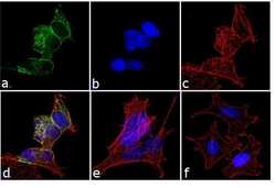
- Immunofluorescence analysis of Phospho-Huntingtin/Htt pSer421 was performed using 70% confluent log phase SH-SY5Y cells which were serum starved (48 h) and stimulated with 50 ng/mL IGF-1 for 30 min. The cells were fixed with 4% paraformaldehyde for 10 minutes, permeabilized with 0.1% Triton™ X-100 for 10 minutes, and blocked with 1% BSA for 1 hour at room temperature. The cells were labeled with Phospho-Huntingtin/Htt pSer421 Rabbit Polyclonal Antibody (Product # PA1-087) at 1:250 dilution in0.1% BSA and incubated for 3 hours at room temperature and then labeled with Goat anti-Rabbit IgG (H+L) Superclonal™ Secondary Antibody, Alexa Fluor® 488 conjugate (Product # A27034) at a dilution of 1:2000 for 45 minutes at room temperature (Panel a: green). Nuclei (Panel b: blue) were stained with SlowFade® Gold Antifade Mountant with DAPI (Product # S36938). F-actin (Panel c: red) was stained with Rhodamine Phalloidin (Product # R415, 1:300). Panel d represents the merged image showing cytoplasmic localization. Panel e shows the untreated cells. Panel f shows the control without primary antibody. The images were captured at 60X magnification.
