Explore
 Validate
 Learn
Learn Western blot
Antibody data
- Antibody Data
- Antigen structure
- References [0]
- Comments [0]
- Validations
- Western blot [2]
- Immunocytochemistry [2]
- Immunohistochemistry [1]
Submit
Validation data
Reference
Comment
Report error
- Product number
- PA5-21586 - Provider product page

- Provider
- Invitrogen Antibodies
- Product name
- HSD11B1 Polyclonal Antibody
- Antibody type
- Polyclonal
- Antigen
- Synthetic peptide
- Description
- Recommended positive controls: HepG2.
- Concentration
- 1 mg/mL
No comments: Submit comment
Supportive validation
- Submitted by
- Invitrogen Antibodies (provider)
- Main image

- Experimental details
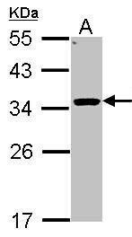
- Western Blot using HSD11B1 Polyclonal Antibody (Product # PA5-21586). Sample (30 µg of whole cell lysate). Lane A: Hep G2. 12% SDS PAGE. HSD11B1 Polyclonal Antibody (Product # PA5-21586) diluted at 1:1,000.
- Submitted by
- Invitrogen Antibodies (provider)
- Main image
- Experimental details
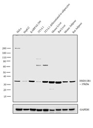
- Western blot analysis was performed on membrane enriched cell extracts (30 µg lysate) of HeLa (Lane 1), HepG2 (Lane 2), KARPAS 299 (Lane 3), 3T3-L1 (Lane 4), 3T3-L1 differentiated to adipocytes (Lane 5), Mouse Liver (Lane 6), Rat Liver (Lane 7), Mouse Adipose (Lane 8) and Rat Adipose (Lane 9). The blot was probed with Anti-HSD11B1 Polyclonal Antibody (Product # PA5-21586, 1:1000 dilution) and detected by chemiluminescence using Goat anti-Rabbit IgG (H+L) Superclonal™ Secondary Antibody, HRP conjugate (Product # A27036, 0.25 µg/ml, 1:4000 dilution). A 35 kDa band corresponding to HSD11B1 was observed in all cell lines and tissues tested and was induced upon differentiation of 3T3-L1 cells to adipocytes.
Supportive validation
- Submitted by
- Invitrogen Antibodies (provider)
- Main image
- Experimental details
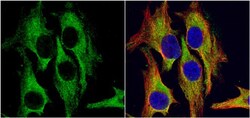
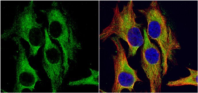
- Immunocytochemistry-Immunofluorescence analysis of HSD11B1 was performed in HeLa cells fixed in 4% paraformaldehyde at RT for 15 min. Green: HSD11B1 Polyclonal Antibody (Product # PA5-21586) diluted at 1:500. Red: alpha Tubulin, a cytoskeleton marker. Blue: Hoechst 33342 staining.
- Submitted by
- Invitrogen Antibodies (provider)
- Main image
- Experimental details
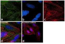
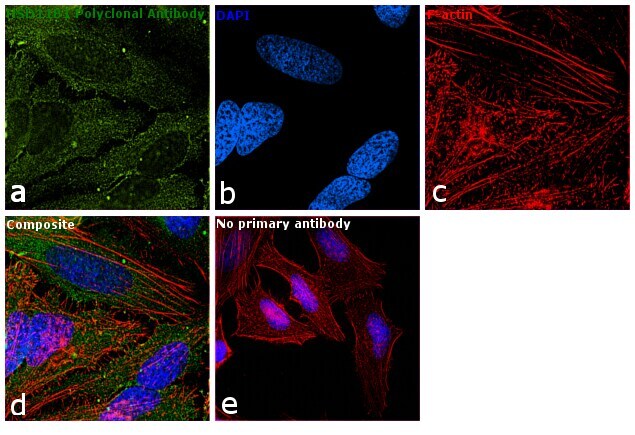
- Immunofluorescence analysis of HSD11B1 was performed using 70% confluent log phase HeLa cells. The cells were fixed with 4% paraformaldehyde for 10 minutes, permeabilized with 0.1% Triton™ X-100 for 15 minutes, and blocked with 1% BSA for 1 hour at room temperature. The cells were labeled with HSD11B1 Polyclonal Antibody (Product # PA5-21586) at 1:100 dilution in 0.1% BSA, incubated at 4 degree Celsius overnight and then labeled with Goat anti-Rabbit IgG (H+L) Superclonal™ Secondary Antibody, Alexa Fluor® 488 conjugate (Product # A27034) at a dilution of 1:2000 for 45 minutes at room temperature (Panel a: green). Nuclei (Panel b: blue) were stained with ProLong™ Diamond Antifade Mountant with DAPI (Product # P36962). F-actin (Panel c: red) was stained with Rhodamine Phalloidin (Product # R415). Panel d represents the merged image showing cytoplasmic localization. Panel e represents control cells with no primary antibody to assess background. The images were captured at 60X magnification.
Supportive validation
- Submitted by
- Invitrogen Antibodies (provider)
- Main image
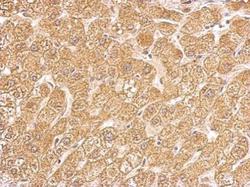
- Experimental details
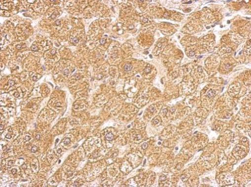
- HSD11B1 Polyclonal Antibody detects HSD11B1 protein at cytosol on human hepatoma by immunohistochemical analysis. Sample: Paraffin-embedded hepatoma. HSD11B1 Polyclonal Antibody (Product # PA5-21586) dilution: 1:500. Antigen Retrieval: EDTA based buffer, pH 8.0, 15 min.