Explore
 Validate
 Learn
Learn Western blot
 Immunohistochemistry
Antibody data
- Antibody Data
- Antigen structure
- References [29]
- Comments [0]
- Validations
- Immunohistochemistry [1]
Submit
Validation data
Reference
Comment
Report error
- Product number
- AMAb91038 - Provider product page

- Provider
- Atlas Antibodies
- Proper citation
- Atlas Antibodies Cat#AMAb91038, RRID:AB_2665776
- Product name
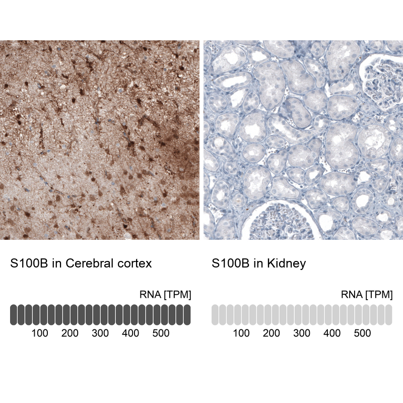
- Anti-S100B
- Antibody type
- Monoclonal
- Description
- Monoclonal Antibody against Human S100B, Clone ID: CL2720, Gene description: S100 calcium binding protein B, Alternative Gene Names: S100beta, Validated applications: IHC, WB, Uniprot ID: P04271, Storage: Store at +4°C for short term storage. Long time storage is recommended at -20°C.
- Reactivity
- Human, Mouse, Rat
- Host
- Mouse
- Conjugate
- Unconjugated
- Isotype
- IgG
- Antibody clone number
- CL2720
- Vial size
- 100 µl
- Concentration
- 1.0 mg/ml
- Storage
- Store at +4°C for short term storage. Long time storage is recommended at -20°C.
- Handling
- The antibody solution should be gently mixed before use.
Submitted references Functional Regrowth of Norepinephrine Axons in the Adult Mouse Brain Following Injury
Cell-Type-Specific Expression of Leptin Receptors in the Mouse Forebrain
Brain organoids engineered to give rise to glia and neural networks after 90 days in culture exhibit human-specific proteoforms.
Upregulation of carbonic anhydrase 1 beneficial for depressive disorder
Functional Cooperation of α-Synuclein and Tau Is Essential for Proper Corticogenesis
Boosting the peripheral immune response in the skeletal muscles improved motor function in ALS transgenic mice
AgRP neurons control structure and function of the medial prefrontal cortex
Inhibition of Schwann cell pannexin 1 attenuates neuropathic pain through the suppression of inflammatory responses
The smoothened agonist SAG reduces mitochondrial dysfunction and neurotoxicity of frataxin-deficient astrocytes
Dihuang-Yinzi Alleviates Cognition Deficits via Targeting Energy-Related Metabolism in an Alzheimer Mouse Model as Demonstrated by Integration of Metabolomics and Network Pharmacology.
Conditioned Medium From the Stem Cells of Human Exfoliated Deciduous Teeth Ameliorates Neuropathic Pain in a Partial Sciatic Nerve Ligation Model
Dedifferentiated Schwann cell-derived TGF-β3 is essential for the neural system to promote wound healing.
Beyond New Neurons in the Adult Hippocampus: Imipramine Acts as a Pro-Astrogliogenic Factor and Rescues Cognitive Impairments Induced by Stress Exposure
BDNF-dependent modulation of axonal transport is selectively impaired in ALS.
Interactions between Autophagy, Proinflammatory Cytokines, and Apoptosis in Neuropathic Pain: Granulocyte Colony Stimulating Factor as a Multipotent Therapy in Rats with Chronic Constriction Injury
Age-related changes in brain phospholipids and bioactive lipids in the APP knock-in mouse model of Alzheimer’s disease
Reduced astrocytic reactivity in human brains and midbrain organoids with PRKN mutations
Neuron-Derived Estrogen Is Critical for Astrocyte Activation and Neuroprotection of the Ischemic Brain
Persistent Cyfip1 Expression Is Required to Maintain the Adult Subventricular Zone Neurogenic Niche
Ependyma‐expressed CCN 1 restricts the size of the neural stem cell pool in the adult ventricular‐subventricular zone
Deletion of astrocytic BMAL1 results in metabolic imbalance and shorter lifespan in mice
Bruton’s Tyrosine Kinase Inhibition Promotes Myelin Repair
APOE4 Causes Widespread Molecular and Cellular Alterations Associated with Alzheimer’s Disease Phenotypes in Human iPSC-Derived Brain Cell Types
Presymptomatic change in microRNAs modulates Tau pathology
Expression of nerve growth factor carried by pseudotyped lentivirus improves neuron survival and cognitive functional recovery of post‐ischemia in rats
Schwann cell–derived periostin promotes autoimmune peripheral polyneuropathy via macrophage recruitment
Reactive Astrocytes Promote ALS-like Degeneration and Intracellular Protein Aggregation in Human Motor Neurons by Disrupting Autophagy through TGF-β1
Astrocyte deletion of Bmal1 alters daily locomotor activity and cognitive functions via GABA signalling
Cooke P, Linden D
eneuro 2025;12(1):ENEURO.0418-24.2024
eneuro 2025;12(1):ENEURO.0418-24.2024
Cell-Type-Specific Expression of Leptin Receptors in the Mouse Forebrain
Canepa C, Kara J, Lee C
International Journal of Molecular Sciences 2024;25(18):9854
International Journal of Molecular Sciences 2024;25(18):9854
Brain organoids engineered to give rise to glia and neural networks after 90 days in culture exhibit human-specific proteoforms.
Wenzel TJ, Mousseau DD
Frontiers in cellular neuroscience 2024;18:1383688
Frontiers in cellular neuroscience 2024;18:1383688
Palau F, Cantarero L, Roldán M, Rodríguez-Sanz M, Mathison A, Díaz-Osorio Y, Pijuan J, Frías M, Urrutia R, Hoenicka J
2024
2024
Upregulation of carbonic anhydrase 1 beneficial for depressive disorder
Cheng K, Wang Y, He Y, Tian Y, Li J, Chen C, Xu X, Wu Z, Yu H, Chen X, Wu Y, Song W, Dong Z, Xu H, Xie P
Acta Neuropathologica Communications 2023;11(1)
Acta Neuropathologica Communications 2023;11(1)
Functional Cooperation of α-Synuclein and Tau Is Essential for Proper Corticogenesis
Wang S, Fu Y, Miyata T, Matsumoto S, Shinoda T, Itoh K, Harada A, Hirotsune S, Jin M
The Journal of Neuroscience 2022;42(37):7031-7046
The Journal of Neuroscience 2022;42(37):7031-7046
Boosting the peripheral immune response in the skeletal muscles improved motor function in ALS transgenic mice
Trolese M, Scarpa C, Melfi V, Fabbrizio P, Sironi F, Rossi M, Bendotti C, Nardo G
Molecular Therapy 2022;30(8):2760-2784
Molecular Therapy 2022;30(8):2760-2784
AgRP neurons control structure and function of the medial prefrontal cortex
Stutz B, Waterson M, Šestan-Peša M, Dietrich M, Škarica M, Sestan N, Racz B, Magyar A, Sotonyi P, Liu Z, Gao X, Matyas F, Stoiljkovic M, Horvath T
Molecular Psychiatry 2022;27(10):3951-3960
Molecular Psychiatry 2022;27(10):3951-3960
Inhibition of Schwann cell pannexin 1 attenuates neuropathic pain through the suppression of inflammatory responses
Wang Q, Li H, Ling Z, Chen G, Wei Z
Journal of Neuroinflammation 2022;19(1)
Journal of Neuroinflammation 2022;19(1)
The smoothened agonist SAG reduces mitochondrial dysfunction and neurotoxicity of frataxin-deficient astrocytes
Vicente-Acosta A, Giménez-Cassina A, Díaz-Nido J, Loria F
Journal of Neuroinflammation 2022;19(1)
Journal of Neuroinflammation 2022;19(1)
Dihuang-Yinzi Alleviates Cognition Deficits via Targeting Energy-Related Metabolism in an Alzheimer Mouse Model as Demonstrated by Integration of Metabolomics and Network Pharmacology.
Han G, Zhen W, Dai Y, Yu H, Li D, Ma T
Frontiers in aging neuroscience 2022;14:873929
Frontiers in aging neuroscience 2022;14:873929
Conditioned Medium From the Stem Cells of Human Exfoliated Deciduous Teeth Ameliorates Neuropathic Pain in a Partial Sciatic Nerve Ligation Model
Liu Y, Kano F, Hashimoto N, Xia L, Zhou Q, Feng X, Hibi H, Miyazaki A, Iwamoto T, Matsuka Y, Zhang Z, Tanaka E, Yamamoto A
Frontiers in Pharmacology 2022;13
Frontiers in Pharmacology 2022;13
Dedifferentiated Schwann cell-derived TGF-β3 is essential for the neural system to promote wound healing.
Ou MY, Tan PC, Xie Y, Liu K, Gao YM, Yang XS, Zhou SB, Li QF
Theranostics 2022;12(12):5470-5487
Theranostics 2022;12(12):5470-5487
Beyond New Neurons in the Adult Hippocampus: Imipramine Acts as a Pro-Astrogliogenic Factor and Rescues Cognitive Impairments Induced by Stress Exposure
Machado-Santos A, Loureiro-Campos E, Patrício P, Araújo B, Alves N, Mateus-Pinheiro A, Correia J, Morais M, Bessa J, Sousa N, Rodrigues A, Oliveira J, Pinto L
Cells 2022;11(3):390
Cells 2022;11(3):390
BDNF-dependent modulation of axonal transport is selectively impaired in ALS.
Tosolini AP, Sleigh JN, Surana S, Rhymes ER, Cahalan SD, Schiavo G
Acta neuropathologica communications 2022 Aug 22;10(1):121
Acta neuropathologica communications 2022 Aug 22;10(1):121
Interactions between Autophagy, Proinflammatory Cytokines, and Apoptosis in Neuropathic Pain: Granulocyte Colony Stimulating Factor as a Multipotent Therapy in Rats with Chronic Constriction Injury
Liao M, Yeh S, Lu K, Hsu J, Chao P, Hsu H, Peng C, Lee Y, Hung Y, Ro L
Biomedicines 2021;9(5):542
Biomedicines 2021;9(5):542
Age-related changes in brain phospholipids and bioactive lipids in the APP knock-in mouse model of Alzheimer’s disease
Emre C, Do K, Jun B, Hjorth E, Alcalde S, Kautzmann M, Gordon W, Nilsson P, Bazan N, Schultzberg M
Acta Neuropathologica Communications 2021;9(1)
Acta Neuropathologica Communications 2021;9(1)
Reduced astrocytic reactivity in human brains and midbrain organoids with PRKN mutations
Kano M, Takanashi M, Oyama G, Yoritaka A, Hatano T, Shiba-Fukushima K, Nagai M, Nishiyama K, Hasegawa K, Inoshita T, Ishikawa K, Akamatsu W, Imai Y, Bolognin S, Schwamborn J, Hattori N
npj Parkinson's Disease 2020;6(1)
npj Parkinson's Disease 2020;6(1)
Neuron-Derived Estrogen Is Critical for Astrocyte Activation and Neuroprotection of the Ischemic Brain
Lu Y, Sareddy G, Wang J, Zhang Q, Tang F, Pratap U, Tekmal R, Vadlamudi R, Brann D
The Journal of Neuroscience 2020;40(38):7355-7374
The Journal of Neuroscience 2020;40(38):7355-7374
Persistent Cyfip1 Expression Is Required to Maintain the Adult Subventricular Zone Neurogenic Niche
Habela C, Yoon K, Kim N, Taga A, Bell K, Bergles D, Maragakis N, Ming G, Song H
The Journal of Neuroscience 2020;40(10):2015-2024
The Journal of Neuroscience 2020;40(10):2015-2024
Ependyma‐expressed CCN 1 restricts the size of the neural stem cell pool in the adult ventricular‐subventricular zone
Wu J, Tian W, Liu Y, Wang H, Zheng J, Wang X, Pan H, Li J, Luo J, Yang X, Lau L, Ghashghaei H, Shen Q
The EMBO Journal 2020;39(5)
The EMBO Journal 2020;39(5)
Deletion of astrocytic BMAL1 results in metabolic imbalance and shorter lifespan in mice
Barca‐Mayo O, Boender A, Armirotti A, De Pietri Tonelli D
Glia 2019;68(6):1131-1147
Glia 2019;68(6):1131-1147
Bruton’s Tyrosine Kinase Inhibition Promotes Myelin Repair
Martin E, Aigrot M, Grenningloh R, Stankoff B, Lubetzki C, Boschert U, Zalc B
Brain Plasticity 2019;5(2):123-133
Brain Plasticity 2019;5(2):123-133
APOE4 Causes Widespread Molecular and Cellular Alterations Associated with Alzheimer’s Disease Phenotypes in Human iPSC-Derived Brain Cell Types
Lin Y, Seo J, Gao F, Feldman H, Wen H, Penney J, Cam H, Gjoneska E, Raja W, Cheng J, Rueda R, Kritskiy O, Abdurrob F, Peng Z, Milo B, Yu C, Elmsaouri S, Dey D, Ko T, Yankner B, Tsai L
Neuron 2018;98(6):1141-1154.e7
Neuron 2018;98(6):1141-1154.e7
Presymptomatic change in microRNAs modulates Tau pathology
Sharma S, Khadimallah I, Corya A, Ali Y, Rao X, Liu Y, Lu H
Scientific Reports 2018;8(1)
Scientific Reports 2018;8(1)
Expression of nerve growth factor carried by pseudotyped lentivirus improves neuron survival and cognitive functional recovery of post‐ischemia in rats
Cao J, Lin Y, Han Y, Ding S, Fan Y, Pan Y, Zhao B, Guo Q, Sun W, Wan J, Tong X
CNS Neuroscience & Therapeutics 2018;24(6):508-518
CNS Neuroscience & Therapeutics 2018;24(6):508-518
Schwann cell–derived periostin promotes autoimmune peripheral polyneuropathy via macrophage recruitment
Allard D, Wang Y, Li J, Conley B, Xu E, Sailer D, Kimpston C, Notini R, Smith C, Koseoglu E, Starmer J, Zeng X, Howard J, Hoke A, Scherer S, Su M
Journal of Clinical Investigation 2018;128(10):4727-4741
Journal of Clinical Investigation 2018;128(10):4727-4741
Reactive Astrocytes Promote ALS-like Degeneration and Intracellular Protein Aggregation in Human Motor Neurons by Disrupting Autophagy through TGF-β1
Tripathi P, Rodriguez-Muela N, Klim J, de Boer A, Agrawal S, Sandoe J, Lopes C, Ogliari K, Williams L, Shear M, Rubin L, Eggan K, Zhou Q
Stem Cell Reports 2017;9(2):667-680
Stem Cell Reports 2017;9(2):667-680
Astrocyte deletion of Bmal1 alters daily locomotor activity and cognitive functions via GABA signalling
Barca-Mayo O, Pons-Espinal M, Follert P, Armirotti A, Berdondini L, De Pietri Tonelli D
Nature Communications 2017;8(1)
Nature Communications 2017;8(1)
No comments: Submit comment
Supportive validation
- Submitted by
- Atlas Antibodies (provider)
- Enhanced method
- Orthogonal validation
- Main image
- Experimental details
- Immunohistochemistry analysis in human cerebral cortex and kidney tissues using AMAb91038 antibody. Corresponding S100B RNA-seq data are presented for the same tissues.
- Sample type
- Human
- Protocol
- Protocol