Explore
 Validate
 Learn
Learn Western blot
Antibody data
- Antibody Data
- Antigen structure
- References [1]
- Comments [0]
- Validations
- Western blot [1]
- Immunocytochemistry [2]
- Immunohistochemistry [4]
- Flow cytometry [7]
Submit
Validation data
Reference
Comment
Report error
- Product number
- NBP2-45224 - Provider product page

- Provider
- Novus Biologicals
- Product name
- Mouse Monoclonal S100B Antibody
- Antibody type
- Monoclonal
- Description
- Protein G purified.
- Reactivity
- Human, Rat
- Host
- Mouse
- Isotype
- IgG
- Vial size
- 0.1 mg
- Concentration
- 1.0 mg/ml
- Storage
- Store at 4C short term. Aliquot and store at -20C long term. Avoid freeze-thaw cycles.
Submitted references Long-term electrical stimulation at ear and electro-acupuncture at ST36-ST37 attenuated COX-2 in the CA1 of hippocampus in kainic acid-induced epileptic seizure rats.
Liao ET, Tang NY, Lin YW, Liang Hsieh C
Scientific reports 2017 Mar 28;7(1):472
Scientific reports 2017 Mar 28;7(1):472
No comments: Submit comment
Supportive validation
- Submitted by
- Novus Biologicals (provider)
- Main image
- Experimental details
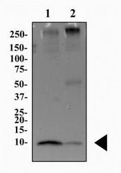
- Western Blot: S100B Antibody (15F4NB) [NBP2-45224] - Total protein from human brain (lane 1) and mouse brain (lane 2) was separated on a 4-15% gel by SDS-PAGE, transferred to 0.2 um PVDF membrane for 30 minutes and blocked in 5% non-fat milk in TBST. The membrane was probed with 2 ug/ml anti-S100B in 1% milk and detected with an anti-mouse HRP secondary antibody using chemiluminescence.

Supportive validation
- Submitted by
- Novus Biologicals (provider)
- Main image
- Experimental details
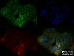
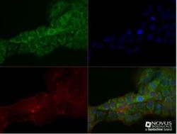
- Immunocytochemistry/Immunofluorescence: S100B Antibody (15F4NB) [NBP2-45224] - A431 cells were fixed for 10 minutes using 10% formalin and then permeabilized for 5 minutes using 1X TBS + 0.5% Triton-X100. The cells were incubated with S100B (15F4NB) at a 1:100 dilution overnight at 4 degrees Celsius and detected with Dylight 488 (Green) at a 1:500 dilution. Actin was detected with Phalloidin 568 (Red) at a 1:200 dilution. Nuclei were detected with DAPI (Blue) at 2.0 ug/ml in 1X PBS. Cells were imaged using a 40X objective.
- Submitted by
- Novus Biologicals (provider)
- Main image
- Experimental details
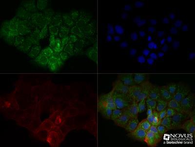
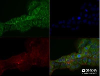
- Immunocytochemistry/Immunofluorescence: S100B Antibody (15F4NB) [NBP2-45224] - A431 cells were fixed for 10 minutes using 10% formalin and then permeabilized for 5 minutes using 1X TBS + 0.5% Triton-X100. The cells were incubated with S100B (15F9NB) at a 1:100 dilution overnight at 4 degrees Celsius and detected with Dylight 488 (Green) at a 1:500 dilution. Actin was detected with Phalloidin 568 (Red) at a 1:200 dilution. Nuclei were detected with DAPI (Blue) at 2.0 ug/ml in 1X PBS. Cells were imaged using a 40X objective. Image using the Azide Free form of this antibody.
Supportive validation
- Submitted by
- Novus Biologicals (provider)
- Main image
- Experimental details
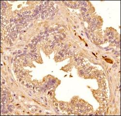
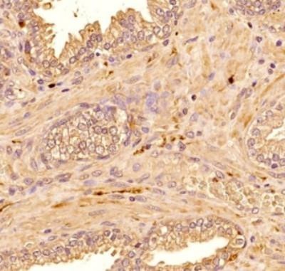
- Immunohistochemistry-Paraffin: S100B Antibody (15F4NB) [NBP2-45224] - IHC analysis of S100B (15F4NB) in human prostate tissue.

- Submitted by
- Novus Biologicals (provider)
- Main image
- Experimental details
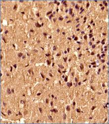
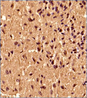
- Immunohistochemistry-Paraffin: S100B Antibody (15F4NB) [NBP2-45224] - IHC analysis of S100B (15F4BN) in human brain tissue.
- Submitted by
- Novus Biologicals (provider)
- Main image

- Experimental details
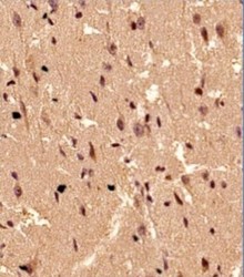
- Immunohistochemistry-Paraffin: S100B Antibody (15F4NB) [NBP2-45224] - Analysis using Azide Free version of NBP2-45224. IHC S100B in human prostate
- Submitted by
- Novus Biologicals (provider)
- Main image
- Experimental details
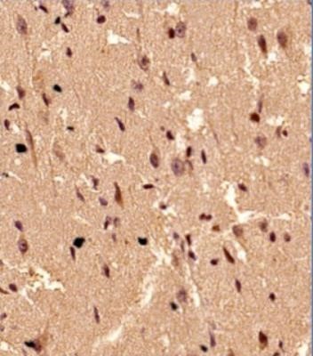
- Immunohistochemistry: S100B Antibody (15F4NB) [NBP2-45224] - Analysis using Azide Free version of NBP2-45224. IHC S100B (15F9NB) in human brain.
Supportive validation
- Submitted by
- Novus Biologicals (provider)
- Main image
- Experimental details
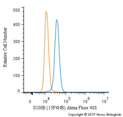
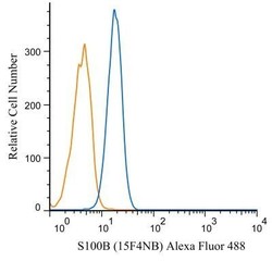
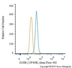
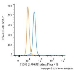
- Flow Cytometry: S100B Antibody (15F4NB) [NBP2-45224] - An intracellular stain was performed on Jurkat cells with S100b (15F4NB) antibody NBP2-45224AF488 (blue) and a matched isotype control (orange). Cells were fixed with 4% PFA and then permeablized with 0.1% saponin. Cells were incubated in an antibody dilution of 5 ug/mL for 30 minutes at room temperature. Both antibodies were conjugated to Alexa Fluor 488.
- Submitted by
- Novus Biologicals (provider)
- Main image
- Experimental details
- Flow Cytometry: S100B Antibody (15F4NB) [NBP2-45224] - Analysis using the Azide Free version of NBP2-45224. Staining of A431 cells (1 x 10^6 cells/ml) with S100B antibody, clone 15F4.1D2 (orange) stained at a dilution of 1:500. Detected with a GtxRb Dylight 488 secondary. Shown with the secondary control (blue).
- Submitted by
- Novus Biologicals (provider)
- Main image
- Experimental details
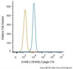
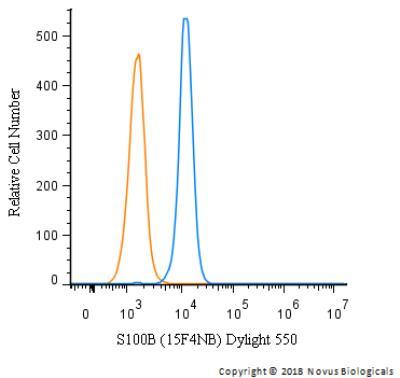
- Flow Cytometry: S100B Antibody (15F4NB) [NBP2-45224] - An intracellular stain was performed on SK-MEL-28 cells with S100b (15F4NB) antibody NBP2-45224R (blue) and a matched isotype control (orange). Cells were fixed with 4% PFA and then permeablized with 0.1% saponin. Cells were incubated in an antibody dilution of 5 ug/mL for 30 minutes at room temperature. Both antibodies were conjugated to Dylight 550.
- Submitted by
- Novus Biologicals (provider)
- Main image
- Experimental details
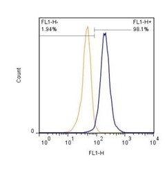
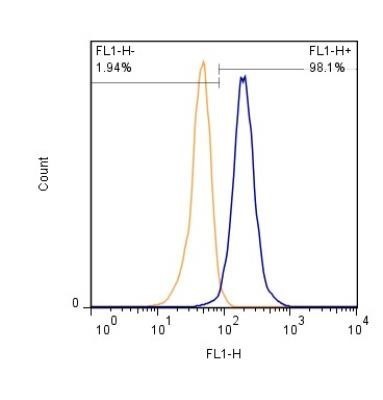
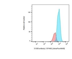
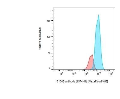
- Flow Cytometry: S100B Antibody (15F4NB) [NBP2-45224] - FHuman primary astrocytes. Histogram overlay of unstained control (in red), and stained primary astrocytes (in blue). Image from verified customer review.
- Submitted by
- Novus Biologicals (provider)
- Main image
- Experimental details
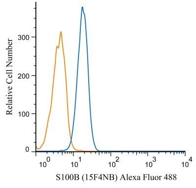
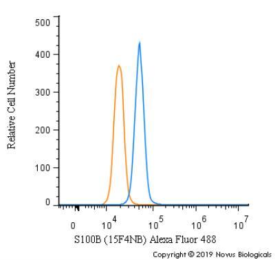
- Flow Cytometry: S100B Antibody (15F4NB) [NBP2-45224] - An intracellular stain was performed on SK-MEL-28 cells with S100B [15F4NB] Antibody NBP2-45224AF488 (blue) and a matched isotype control (orange). Cells were fixed with 4% PFA and then permeabilized with 0.1% saponin. Cells were incubated in an antibody dilution of 5 ug/mL for 30 minutes at room temperature. Both antibodies were conjugated to Alexa Fluor 488.
- Submitted by
- Novus Biologicals (provider)
- Main image
- Experimental details
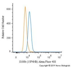
- Flow Cytometry: S100B Antibody (15F4NB) [NBP2-45224] - An intracellular stain was performed on A431 cells with S100b [15F4NB] Antibody NBP2-45224AF488 (blue) and a matched isotype control (orange). Cells were fixed with 4% PFA and then permeabilized with 0.1% saponin. Cells were incubated in an antibody dilution of 5 ug/mL for 30 minutes at room temperature. Both antibodies were conjugated to Alexa Fluor 488.
- Submitted by
- Novus Biologicals (provider)
- Main image
- Experimental details
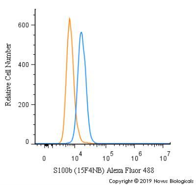
- Flow Cytometry: S100B Antibody (15F4NB) [NBP2-45224] - An intracellular stain was performed on SK-MEL-28 cells with S100b [15F4NB] Antibody NBP2-45224AF488 (blue) and a matched isotype control (orange). Cells were fixed with 4% PFA and then permeabilized with 0.1% saponin. Cells were incubated in an antibody dilution of 5 ug/mL for 30 minutes at room temperature. Both antibodies were conjugated to Alexa Fluor 488.