Explore
 Validate
 Learn
Learn Western blot
 Immunocytochemistry
Antibody data
- Antibody Data
- Antigen structure
- References [3]
- Comments [0]
- Validations
- Immunocytochemistry [2]
- Immunohistochemistry [4]
- Other assay [3]
Submit
Validation data
Reference
Comment
Report error
- Product number
- PA5-78161 - Provider product page

- Provider
- Invitrogen Antibodies
- Product name
- S100B Polyclonal Antibody
- Antibody type
- Polyclonal
- Antigen
- Recombinant full-length protein
- Description
- Positive Control: V5-human S100B-transfected 293T, A375, mouse brain, rat brain Predicted Reactivity: Rabbit (100%), Chicken (84%), Rhesus Monkey (98%), Bovine (96%) Store product as a concentrated solution. Centrifuge briefly prior to opening the vial.
- Reactivity
- Human, Mouse, Rat
- Host
- Rabbit
- Isotype
- IgG
- Vial size
- 100 μL
- Concentration
- 0.14 mg/mL
- Storage
- Store at 4°C short term. For long term storage, store at -20°C, avoiding freeze/thaw cycles.
Submitted references Novel patient-derived xenograft and cell line models for therapeutic screening in NF2-associated schwannoma.
Murine muscle stem cell response to perturbations of the neuromuscular junction are attenuated with aging.
Oligodendrocytes are susceptible to Zika virus infection in a mouse model of perinatal exposure: Implications for CNS complications.
Zhao F, Chen Y, Li SW, Zhang J, Zhang S, Zhao XB, Yang ZJ, Wang B, He QY, Wang LM, Xu L, Liu PN
The Journal of pathology 2022 Aug;257(5):620-634
The Journal of pathology 2022 Aug;257(5):620-634
Murine muscle stem cell response to perturbations of the neuromuscular junction are attenuated with aging.
Larouche JA, Mohiuddin M, Choi JJ, Ulintz PJ, Fraczek P, Sabin K, Pitchiaya S, Kurpiers SJ, Castor-Macias J, Liu W, Hastings RL, Brown LA, Markworth JF, De Silva K, Levi B, Merajver SD, Valdez G, Chakkalakal JV, Jang YC, Brooks SV, Aguilar CA
eLife 2021 Jul 29;10
eLife 2021 Jul 29;10
Oligodendrocytes are susceptible to Zika virus infection in a mouse model of perinatal exposure: Implications for CNS complications.
Schultz V, Barrie JA, Donald CL, Crawford CL, Mullin M, Anderson TJ, Solomon T, Barnett SC, Linington C, Kohl A, Willison HJ, Edgar JM
Glia 2021 Aug;69(8):2023-2036
Glia 2021 Aug;69(8):2023-2036
No comments: Submit comment
Supportive validation
- Submitted by
- Invitrogen Antibodies (provider)
- Main image
- Experimental details
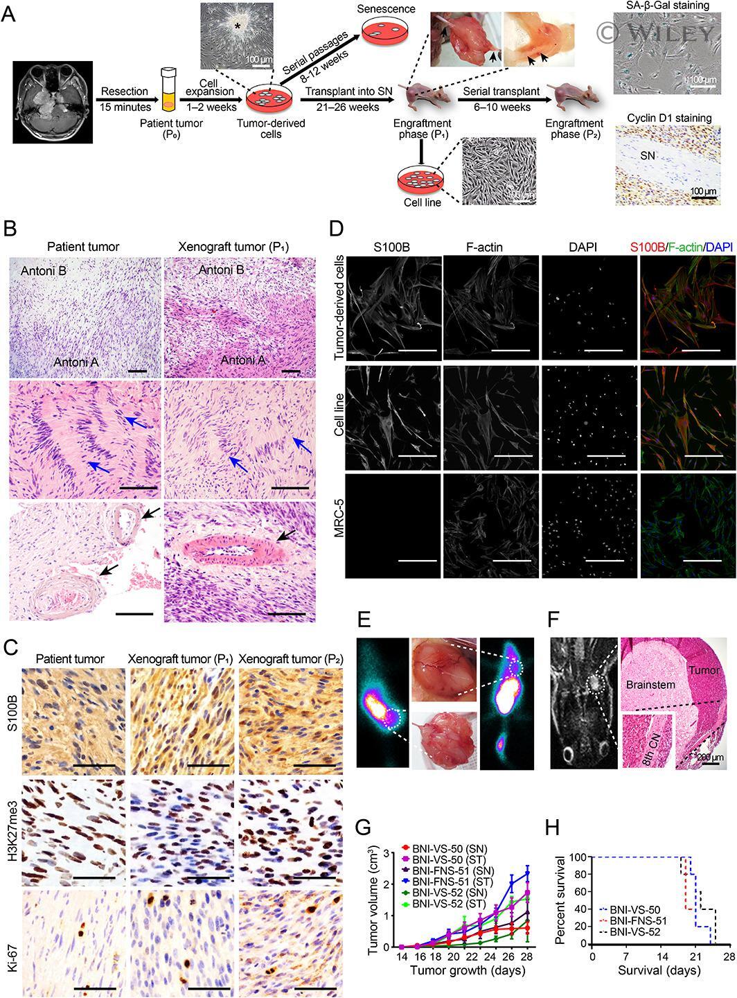
- S100B Polyclonal Antibody detects S100 beta protein at glia cells by immunofluorescent analysis. Sample: DIV10 rat E18 primary cortical neuron and glia cells were fixed in 4% paraformaldehyde at RT for 15 min. Green: S100 beta stained by S100B Polyclonal Antibody (Product # PA5-78161) diluted at 1:500. Red: Tau, stained by Phospho-Tau (Ser262) Polyclonal Antibody [GT287]diluted at 1:500. Blue: Fluoroshield with DAPI .
- Submitted by
- Invitrogen Antibodies (provider)
- Main image

- Experimental details
- S100B Polyclonal Antibody detects S100 beta protein at glia cells by immunofluorescent analysis. Sample: DIV10 rat E18 primary cortical neuron and glia cells were fixed in 4% paraformaldehyde at RT for 15 min. Green: S100 beta stained by S100B Polyclonal Antibody (Product # PA5-78161) diluted at 1:500. Red: Tau, stained by Phospho-Tau (Ser262) Polyclonal Antibody [GT287]diluted at 1:500. Blue: Fluoroshield with DAPI .

Supportive validation
- Submitted by
- Invitrogen Antibodies (provider)
- Main image
- Experimental details
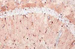
- S100B Polyclonal Antibody detects S100 beta protein at cytoplasm by immunohistochemical analysis. Sample: Paraffin-embedded rat brain. S100 beta stained by S100B Polyclonal Antibody (Product # PA5-78161) diluted at 1:500. Antigen Retrieval: Citrate buffer, pH 6.0, 15 min.
- Submitted by
- Invitrogen Antibodies (provider)
- Main image

- Experimental details
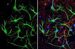
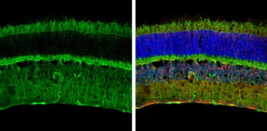
- S100B Polyclonal Antibody detects S100 beta protein expression by immunohistochemical analysis. Sample: Frozen sectioned adult mouse retina. Green: S100 beta stained by S100B Polyclonal Antibody (Product # PA5-78161) diluted at 1:250. Red: beta Tubulin 3/ TUJ1, stained by beta Tubulin 3/ TUJ1 antibody [GT11710] diluted at 1:250. Blue: Fluoroshield with DAPI .
- Submitted by
- Invitrogen Antibodies (provider)
- Main image
- Experimental details
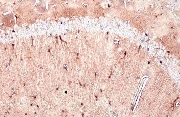
- S100B Polyclonal Antibody detects S100 beta protein at cytoplasm by immunohistochemical analysis. Sample: Paraffin-embedded mouse brain. S100 beta stained by S100B Polyclonal Antibody (Product # PA5-78161) diluted at 1:500. Antigen Retrieval: Citrate buffer, pH 6.0, 15 min.
- Submitted by
- Invitrogen Antibodies (provider)
- Main image
- Experimental details
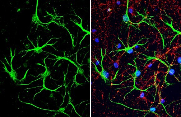
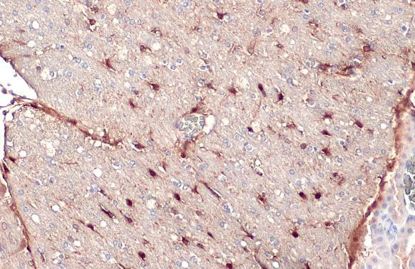
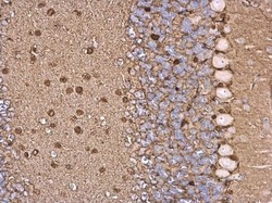
- S100B antibody detects S100B protein at glial cell on mouse hind brain by immunohistochemical analysis. Sample: Paraffin-embedded mouse hind brain. S100B antibody (Product # PA5-78161) dilution: 1:500. Antigen Retrieval: EDTA based buffer, pH 8.0, 15 min.

Supportive validation
- Submitted by
- Invitrogen Antibodies (provider)
- Main image
- Experimental details
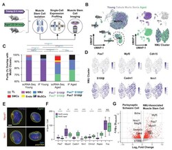
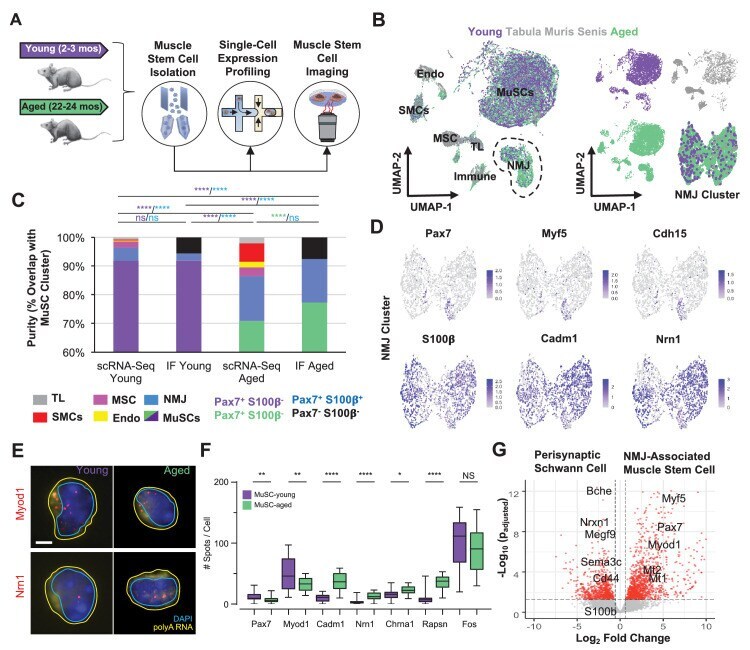
- Figure 3. Single cell analysis of skeletal muscle stem cells (MuSCs) in age shows a subset express transcripts associated with the neuromuscular junction. ( A ) Experiment design schematic. MuSCs were fluorescent activated cell sorting (FACS) enriched from young and aged wild-type (WT) mice, then analyzed by IF, single cell mRNA sequencing (scRNA-Seq), or single-molecule fluorescence in situ hybridization (smFISH). ( B ) Left: Dimensional reduction and unsupervised clustering of young (2-3 months) and aged (22-24 months) WT FACS-isolated MuSCs and limb muscle cells from 24-month Tabula Muris Senis datasets colored by sample type showing overlap between FACS-enriched MuSCs and MuSC clusters among all mono-nucleated cells. Four and five independent replicates of young and age, respectively, were sequenced where each sample is composed of a pool from two mice. Cell types were identified according to markergene expression and labeled on the plot. NMJ - neuromuscular junction; TL - tenocyte; MSC - mesenchymal stromal cell; Immune - CD45+ immune cell; Endo - endothelial cell; SMC - smooth muscle cell. Right: Plots split by sample type showing contribution to uniform manifold approximation and projection (UMAP) and re-clustered UMAP of the NMJ-associated cluster. ( C ) Stacked bar plot representing the fraction of the sorted cell populations that clustered with each mono-nucleated cell type as well as the number of Pax7+S100b- (MuSC) and Pax7+S100b+ (NMJ) cells based on IF stains. A
- Submitted by
- Invitrogen Antibodies (provider)
- Main image

- Experimental details
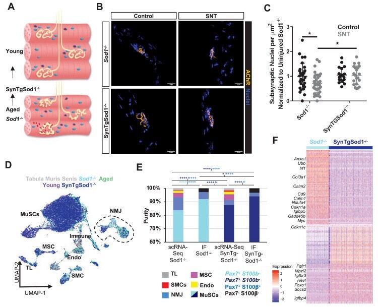
- Figure 5. Neurodegeneration activates muscle stem cells (MuSCs) in a similar manner to aging and rescue of motor neurons partially reverses this activation state. ( A ) Schematic of neuromuscular junction (NMJ) in young (top) and aged or Sod1 -/- (bottom) muscle, whereby NMJ becomes fragmented or partially denervated. ( B ) Representative images of Sod -/- and SynTgSod1 -/- myofibers from uninjured and sciatic nerve transection (SNT) tibialis anterior muscles stained with DAPI (blue) and alpha-bungarotoxin (orange). ( C ) Quantification of the number of subsynaptic nuclei per synaptic area in uninjured control and SNT muscle fibers from both Sod1 -/- and SynTgSod1 -/- mice, normalized to the subsynaptic nuclei per area of Sod1 -/- uninjured control (*p < 0.05, two-way ANOVA with Tukey post hoc test). ( D ) Uniform manifold approximation and projection (UMAP) dimensional reduction of fluorescent activated cell sorting (FACS)-isolated MuSCs (FSMs) from young and aged wild-type (WT) mice, 10-month Sod1 -/- mice, 10-month Sod1 -/- rescue (SynTgSod1 -/- ) mice, and public Tabula Muris Senis limb muscle datasets colored by sample type. One and two independent replicates of Sod1 -/- and Sod1 -/- rescue (SynTgSod1 -/- ) mice, respectively, were sequenced where each sample is composed of a pool from two mice. Cell types were identified according to markergene expression and labeled on the plot. TL - tenocyte; MSC - mesenchymal stromal cell; Immune - CD45+ immune cell; Endo - endotheli
- Submitted by
- Invitrogen Antibodies (provider)
- Main image
- Experimental details
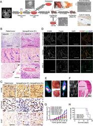
- 1 Figure Morphological and histopathological characterization of PDX and cell line models. (A) Schematic diagram outlining the strategy for the establishment of PDX and cell line models from the patient tumors. Representative images of brain MRI, tumor-derived cells, xenograft in sciatic nerve (SN, black arrow), and cell line illustrating the steps of this protocol are shown. A representative image of tumor-derived cell cluster (asterisk) that augmented the growth of the culture is shown. A representative image of positive staining for beta-galactosidase (SA-beta-Gal) in cultured cells after serial passages for 12 weeks is shown. Representative images of macroscopic tissue and IHC staining for Cyclin D1 are shown. (B) Representative images of HE staining, showing spindle-cell lesions with intermixed (Antoni A) and loose (Antoni B) cellular arrangements, Verocay bodies (blue arrow) and perivascular hyalinization (black arrow) in patient and xenograft tumors. Scale bars, 100 mum. (C) Representative images of IHC staining for S-100, H3K27me3, and Ki-67 in patient and xenograft tumors. Scale bars, 100 mum. (D) Representative images of immunofluorescence staining for S100 and F-actin in tumor-derived cells and cell line. The MRC-5 cell line was used as a negative control for the S100 antibody. DAPI was used to indicate nuclei. Scale bars, 100 mum. (E) Representative images of SPECT imaging, gross appearance, and HE staining, showing the tumor growth after sciatic nerve (left) or s