 Explore
 Validate
 Learn
Learn Western blot
 ELISA
ELISAAntibody data
- Antibody Data
- Antigen structure
- References [1]
- Comments [0]
- Validations
- Western blot [1]
- Immunocytochemistry [2]
- Immunohistochemistry [2]
Submit
Validation data
Reference
Comment
Report error
- Product number
- MA1-751 - Provider product page

- Provider
- Invitrogen Antibodies
- Product name
- Presenilin 1 Monoclonal Antibody (APS 11)
- Antibody type
- Monoclonal
- Antigen
- Synthetic peptide
- Description
- MA1-751 detects presenilin 1 protein (PS1) from mouse, rat, human, and nonhuman primate samples. No cross-reactivity is seen with presenilin 2. MA1-751 has successfully been used immunofluorescence, immunocytochemistry, immunohistochemistry, Western blot , and ELISA procedures. By Western blot, this antibody detects an ~28 kDa protein representing PS1 N-terminus cleavage product from ST15 cell lysate transfected with human PS1. In formalin-fixed, paraffin embedded sections of human brain, this antibody showed strong staining of both the plaque core and dystrophic neurites. The MA1-751 immunogen is a synthetic peptide corresponding to residues C H(21) L S N T V R S Q N D N R E(34) of human PS1.
- Reactivity
- Human, Mouse, Rat
- Host
- Mouse
- Isotype
- IgG
- Antibody clone number
- APS 11
- Vial size
- 200 μg
- Concentration
- 1 mg/mL
- Storage
- -20°C, Avoid Freeze/Thaw Cycles
Submitted references Analysis of presenilin 1 and presenilin 2 expression and processing by newly developed monoclonal antibodies.
Diehlmann A, Ida N, Weggen S, Grünberg J, Haass C, Masters CL, Bayer TA, Beyreuther K
Journal of neuroscience research 1999 May 15;56(4):405-19
Journal of neuroscience research 1999 May 15;56(4):405-19
No comments: Submit comment
Supportive validation
- Submitted by
- Invitrogen Antibodies (provider)
- Main image

- Experimental details
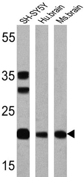
- Western blot analysis of Presenilin 1 was performed by loading 25 µg of SH-SY5Y (lane 1), human brain (lane 2) and mouse brain (lane 3) onto an SDS polyacrylamide gel. Proteins were transferred to a PVDF membrane and blocked at 4ºC overnight. The membrane was probed with a Presenilin 1 monoclonal antibody (Product # MA1-751) at a dilution of 1:200 overnight at 4°C, washed in TBST, and probed with an HRP-conjugated secondary antibody for 1 hr at room temperature in the dark. Chemiluminescent detection was performed using Pierce ECL Plus Western Blotting Substrate (Product # 32132). Images were taken at an exposure time of 2 min (lane 1) and 4 min (lanes 2 and 3). Results show a band at ~22 kDa.
Supportive validation
- Submitted by
- Invitrogen Antibodies (provider)
- Main image
- Experimental details
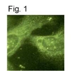
- Immunocytochemical staining of PS1 in mouse fibroblasts using Product # MA1-751.

- Submitted by
- Invitrogen Antibodies (provider)
- Main image

- Experimental details
- Immunocytochemical staining of PS1 in mouse fibroblasts using Product # MA1-751.

Supportive validation
- Submitted by
- Invitrogen Antibodies (provider)
- Main image

- Experimental details
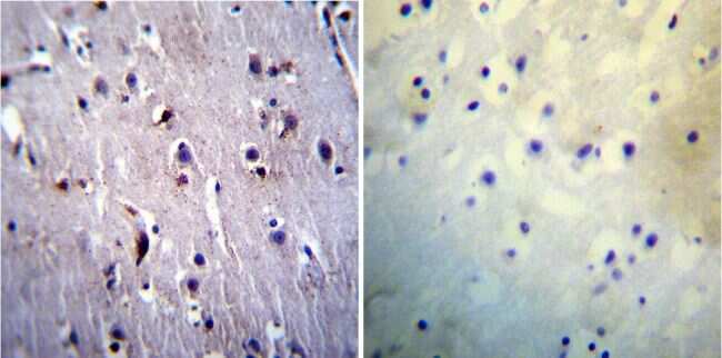
- Immunohistochemistry was performed on normal biopsies of deparaffinized Human brain tissue. To expose target proteins, heat induced antigen retrieval was performed using 10mM sodium citrate (pH6.0) buffer, microwaved for 8-15 minutes. Following antigen retrieval tissues were blocked in 3% BSA-PBS for 30 minutes at room temperature. Tissues were then probed at a dilution of 1:20 with a mouse monoclonal antibody recognizing Presenilin 1 (Product # MA1-751) or without primary antibody (negative control) overnight at 4°C in a humidified chamber. Tissues were washed extensively with PBST and endogenous peroxidase activity was quenched with a peroxidase suppressor. Detection was performed using a biotin-conjugated secondary antibody and SA-HRP, followed by colorimetric detection using DAB. Tissues were counterstained with hematoxylin and prepped for mounting.
- Submitted by
- Invitrogen Antibodies (provider)
- Main image

- Experimental details
- Immunohistochemistry was performed on normal biopsies of deparaffinized Human liver tissue. To expose target proteins, heat induced antigen retrieval was performed using 10mM sodium citrate (pH6.0) buffer, microwaved for 8-15 minutes. Following antigen retrieval tissues were blocked in 3% BSA-PBS for 30 minutes at room temperature. Tissues were then probed at a dilution of 1:200 with a mouse monoclonal antibody recognizing Presenilin 1 (Product # MA1-751) or without primary antibody (negative control) overnight at 4°C in a humidified chamber. Tissues were washed extensively with PBST and endogenous peroxidase activity was quenched with a peroxidase suppressor. Detection was performed using a biotin-conjugated secondary antibody and SA-HRP, followed by colorimetric detection using DAB. Tissues were counterstained with hematoxylin and prepped for mounting.
