 Explore
 Validate
 Learn
Learn Western blot
Antibody data
- Antibody Data
- Antigen structure
- References [1]
- Comments [0]
- Validations
- Western blot [1]
- Immunohistochemistry [9]
Submit
Validation data
Reference
Comment
Report error
- Product number
- NBP2-29623 - Provider product page

- Provider
- Novus Biologicals
- Product name
- Mouse Monoclonal SOX2 Antibody
- Antibody type
- Monoclonal
- Description
- Protein A or G purified.
- Reactivity
- Human
- Host
- Mouse
- Isotype
- IgG
- Vial size
- 0.1 mg
- Concentration
- 1.0 mg/ml
- Storage
- Store at 4C short term. Aliquot and store at -20C long term. Avoid freeze-thaw cycles.
Submitted references Immunohistochemical and Biochemical Expression Patterns of TTF-1, RAGE, GLUT-1 and SOX2 in HCV-Associated Hepatocellular Carcinomas.
Aboushousha T, Mamdouh S, Hamdy H, Helal N, Khorshed F, Safwat G, Seleem M
Asian Pacific journal of cancer prevention : APJCP 2018 Jan 27;19(1):219-227
Asian Pacific journal of cancer prevention : APJCP 2018 Jan 27;19(1):219-227
No comments: Submit comment
Supportive validation
- Submitted by
- Novus Biologicals (provider)
- Main image

- Experimental details
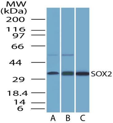
- Western Blot: SOX2 Antibody (4G8) [NBP2-29623] - Western blot detection of SOX2 in A. HEK293 cell lysate B. NCCIT cell lysate and C. Full length recombinant protein.
Supportive validation
- Submitted by
- Novus Biologicals (provider)
- Main image

- Experimental details
- Immunohistochemistry-Paraffin: SOX2 Antibody (4G8) [NBP2-29623] - IHC-P analysis of SOX2 protein in a section of human renal cell carcinoma using 5 ug/ml concentration of SOX2 antibody (clone 4G8). Granular nuclear and cytoplasmic staining pattern was observed in the carcinoma cells, whereas, a weak to negligible staining was observed in the stroma areas [Magnification 40X]..
- Submitted by
- Novus Biologicals (provider)
- Main image

- Experimental details
- Immunohistochemistry-Paraffin: SOX2 Antibody (4G8) [NBP2-29623] - IHC-P analysis of SOX2 protein in a section of human kidney using 5 ug/ml concentration of SOX2 antibody (clone 4G8). The representative image shows distinct nuclear and cytoplasmic staining pattern in cells of different types of renal tubules and blood vessels in the kidney section [Magnification 40X].

- Submitted by
- Novus Biologicals (provider)
- Main image

- Experimental details
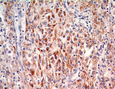
- Immunohistochemistry-Paraffin: SOX2 Antibody (4G8) [NBP2-29623] - IHC analysis of formalin-fixed paraffin-embedded tissue section of normal human liver using mouse monoclonal SOX2 antibody (clone 4G8) at 5 ug/ml concentration. The liver section depicted a diffused cytoplasmic staining in the hepatocytes [Magnification 40X].

- Submitted by
- Novus Biologicals (provider)
- Main image

- Experimental details
- Immunohistochemistry-Paraffin: SOX2 Antibody (4G8) [NBP2-29623] - IHC analysis of formalin-fixed paraffin-embedded tissue section of normal human brain using mouse monoclonal SOX2 antibody (clone 4G8) at 5 ug/ml concentration. The representative photomicrograph depicts a cytoplasmic and nuclear immunopositivity of SOX2 protein in brain [Magnification 40X].

- Submitted by
- Novus Biologicals (provider)
- Main image

- Experimental details
- Immunohistochemistry-Paraffin: SOX2 Antibody (4G8) [NBP2-29623] - IHC analysis of formalin-fixed paraffin-embedded tissue section of human uterine cancer using mouse monoclonal SOX2 antibody (clone 4G8) at 5 ug/ml concentration. The cancer cells developed a punctate staining in the cytoplasm and nuclei, whereas the stroma was very weakly positive for SOX2 protein [Magnification 40X].

- Submitted by
- Novus Biologicals (provider)
- Main image

- Experimental details
- Immunohistochemistry-Paraffin: SOX2 Antibody (4G8) [NBP2-29623] - IHC analysis of formalin-fixed paraffin-embedded tissue section of human lung cancer using mouse monoclonal SOX2 antibody (clone 4G8) at 5 ug/ml concentration. The cancer cells developed a punctate immunopositivity of SOX2 protein in the cytoplasm and the staining was mainly localized in the perinuclear region of the cells [Magnification 40X].

- Submitted by
- Novus Biologicals (provider)
- Main image

- Experimental details
- Immunohistochemistry-Paraffin: SOX2 Antibody (4G8) [NBP2-29623] - IHC analysis of formalin-fixed paraffin-embedded tissue section of human breast cancer using mouse monoclonal SOX2 antibody (clone 4G8) at 5 ug/ml concentration. The cancer cells developed a cytoplasmic and nuclear immunepositivity, whereas the stroma was largely negative for SOX2 protein [Magnification 10X].

- Submitted by
- Novus Biologicals (provider)
- Main image

- Experimental details
- Immunohistochemistry-Paraffin: SOX2 Antibody (4G8) [NBP2-29623] - IHC analysis of formalin-fixed paraffin-embedded tissue section of human bladder cancer using mouse monoclonal SOX2 antibody (clone 4G8) at 5 ug/ml concentration. The cancer cells developed a cytoplasmic and nuclear immunepositivity for SOX2 protein [Magnification 10X].

- Submitted by
- Novus Biologicals (provider)
- Main image

- Experimental details
- Immunohistochemistry-Paraffin: SOX2 Antibody (4G8) [NBP2-29623] - IHC analysis of formalin-fixed paraffin-embedded section of normal human colon tissue using mouse monoclonal SOX2 antibody (clone 4G8) at 5 ug/ml concentration. A weak and diffused cytoplasmic staining was observed in majority of the cells in mucosal layer of colon [Magnification 40X].
