Explore
 Validate
 Learn
Learn Western blot
 Immunoprecipitation
Antibody data
- Antibody Data
- Antigen structure
- References [0]
- Comments [0]
- Validations
- Western blot [1]
- Immunocytochemistry [1]
Submit
Validation data
Reference
Comment
Report error
- Product number
- AM12005PU-N - Provider product page

- Provider
- Acris Antibodies GmbH
- Proper citation
- Acris Antibodies GmbH Cat#AM12005PU-N, RRID:AB_10554868
- Product name
- anti CD3 (activation epitope)
- Antibody type
- Monoclonal
- Antigen
- Purified human CD3 proteins isolated from thymus
- Reactivity
- Human, Mouse
- Host
- Mouse
- Isotype
- IgG
- Antibody clone number
- APA1/1
- Vial size
- 0.1 mg
- Concentration
- 1.0 mg/ml
No comments: Submit comment
Supportive validation
- Submitted by
- Acris Antibodies GmbH (provider)
- Main image
- Experimental details
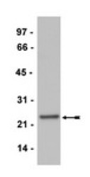
- Jurkat cell lysate was probed with anti-CD3 epsilon AM12005PU-N (0.5μg/ml).

Supportive validation
- Submitted by
- Acris Antibodies GmbH (provider)
- Main image

- Experimental details
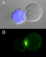
- T-cell Activation by an Antigen presenting Cell. Panel A is an image of a single Antigen-Presenting Cell (APC; a Raji cell, stained blue with CMAC dye) activated by Staphylococcal enterotoxin E in contact with a single T-cell (a Jurkat cell). Panel B is an image of the two cells, stained with an anti-CD3ζ antiserum (a 1:3,000 dilution) and anti-CD3ε, clone APA1/1 (Cat. #AM12005PU-N; 10µg/ml). Visualization of CD3ζ or CD3ε was with an anti-rabbit Alexa 488 (green) or anti-mouse Alexa 594 (red) conjugate, respectively; the yellow color is produced by co-localization of CD3ζ and CD3ε (clone APA1/1) staining. Data generously provided by Dr. Balbino Alarcon, Centro de Biologia Molecular, CSIC, Madrid, Spain.