 Explore
 Validate
 Learn
Learn Immunohistochemistry
 Flow cytometry
Antibody data
- Antibody Data
- Antigen structure
- References [10]
- Comments [0]
- Validations
- Flow cytometry [8]
Submit
Validation data
Reference
Comment
Report error
- Product number
- NBP2-25195 - Provider product page

- Provider
- Novus Biologicals
- Product name
- Mouse Monoclonal CD8 Antibody
- Antibody type
- Monoclonal
Submitted references Glioma-derived extracellular vesicles selectively suppress immune responses.
Preferential elimination of CD28+ T cells in systemic lupus erythematosus (SLE) and the relation with activation-induced apoptosis.
Preferential elimination of CD28+ T cells in systemic lupus erythematosus (SLE) and the relation with activation-induced apoptosis.
CD40 expressed on thymic epithelial cells provides costimulation for proliferation but not for apoptosis of human thymocytes.
Development of retrovirally marked human T progenitor cells into mature thymocytes.
Development of retrovirally marked human T progenitor cells into mature thymocytes.
A monoclonal antibody (A6) recognizing a unique epitope restricted to CD45RO and RB isoforms of the leukocyte common antigen family identifies functional T cell subsets.
Precursors of CD3+CD4+CD8+ cells in the human thymus are defined by expression of CD34. Delineation of early events in human thymic development.
Precursors of CD3+CD4+CD8+ cells in the human thymus are defined by expression of CD34. Delineation of early events in human thymic development.
Immunofluorescent labeling using covalently linked anti-phycoerythrin antibodies and phycoerythrin polymers.
Hellwinkel JE, Redzic JS, Harland TA, Gunaydin D, Anchordoquy TJ, Graner MW
Neuro-oncology 2016 Apr;18(4):497-506
Neuro-oncology 2016 Apr;18(4):497-506
Preferential elimination of CD28+ T cells in systemic lupus erythematosus (SLE) and the relation with activation-induced apoptosis.
Kaneko H, Saito K, Hashimoto H, Yagita H, Okumura K, Azuma M
Clinical and experimental immunology 1996 Nov;106(2):218-29
Clinical and experimental immunology 1996 Nov;106(2):218-29
CD40 expressed on thymic epithelial cells provides costimulation for proliferation but not for apoptosis of human thymocytes.
Ruggiero G, Martinez Cáceres E, Voordouw A, Noteboom E, Graf D, Kroczek RA, Spits H
Journal of immunology (Baltimore, Md. : 1950) 1996 May 15;156(10):3737-46
Journal of immunology (Baltimore, Md. : 1950) 1996 May 15;156(10):3737-46
Development of retrovirally marked human T progenitor cells into mature thymocytes.
Staal FJ, Res PC, Weijer K, Spits H
International immunology 1995 Aug;7(8):1301-9
International immunology 1995 Aug;7(8):1301-9
Development of retrovirally marked human T progenitor cells into mature thymocytes.
Staal FJ, Res PC, Weijer K, Spits H
International immunology 1995 Aug;7(8):1301-9
International immunology 1995 Aug;7(8):1301-9
A monoclonal antibody (A6) recognizing a unique epitope restricted to CD45RO and RB isoforms of the leukocyte common antigen family identifies functional T cell subsets.
Aversa G, Waugh JA, Hall BM
Cellular immunology 1994 Oct 15;158(2):314-28
Cellular immunology 1994 Oct 15;158(2):314-28
Precursors of CD3+CD4+CD8+ cells in the human thymus are defined by expression of CD34. Delineation of early events in human thymic development.
Galy A, Verma S, Bárcena A, Spits H
The Journal of experimental medicine 1993 Aug 1;178(2):391-401
The Journal of experimental medicine 1993 Aug 1;178(2):391-401
Immunofluorescent labeling using covalently linked anti-phycoerythrin antibodies and phycoerythrin polymers.
Wilson MR, Crowley S, Odgers GA, Shaw L
Cytometry 1991;12(4):373-7
Cytometry 1991;12(4):373-7
No comments: Submit comment
Supportive validation
- Submitted by
- Novus Biologicals (provider)
- Main image

- Experimental details
- Flow Cytometry: CD8 alpha Antibody (RPA-T8) [NBP2-25195] - Cell surface flow analysis of CD8 in human PBMC using this antibody at 0.25 ug/10^6 cells. Cells were stained with primary antibody followed by a PE-conjugated goat anti-mouse secondary antibody this antibody . Green represents isotype control this antibody ; red represents anti-CD8 this antibody.

- Submitted by
- Novus Biologicals (provider)
- Main image
- Experimental details
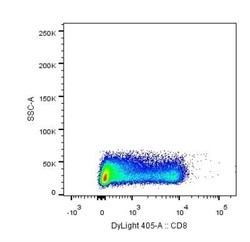
- Flow Cytometry: CD8 alpha Antibody (RPA-T8) [NBP2-25195] - Analysis using the DyLight 405 conjugate of NBP2-25195. Staining of CD8 alpha in human PBMCs using anti-CD8 alpha antibody. Image from verified customer review.

- Submitted by
- Novus Biologicals (provider)
- Main image

- Experimental details
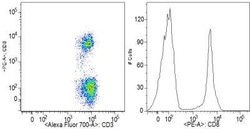
- Flow Cytometry: CD8 alpha Antibody (RPA-T8) [NBP2-25195] - Analysis using the Alexa Fluor (R) 700 conjugate of NBP2-25195. Staining of CD8 alpha in human PBMC using CD8 alpha antibody. Image from verified customer review.

- Submitted by
- Novus Biologicals (provider)
- Main image

- Experimental details
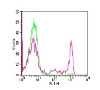
- Flow Cytometry: CD8 alpha Antibody (RPA-T8) [NBP2-25195] - Analysis using the FITC conjugate of NBP2-25195. Staining of CD8 in human PBMCs using 0.25 ug of (clone RPA-T8) per approximately 1x10^6 cells. Green represents isotype control this antibody, red represents anti-CD8 FITC antibody.
- Submitted by
- Novus Biologicals (provider)
- Main image

- Experimental details
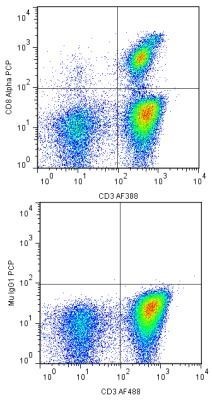
- Flow Cytometry: CD8 alpha Antibody (RPA-T8) [NBP2-25195] - Analysis using PerCP conjugate of NBP2-25195. A cell surface stain was performed on hPBMCs with CD8 Alpha antibody (RPA-T8) NBP2-25195 (top image) and a matched isotype control NBP2-27287 (bottom image). Cells were incubated in an antibody dilution of 1:200 for 20 minutes at room temperature. Both antibodies were conjugated to PerCP. A co-stain was also performed using CD3 antibody NBP2-24867AF488.
- Submitted by
- Novus Biologicals (provider)
- Main image

- Experimental details
- Flow Cytometry: CD8 alpha Antibody (RPA-T8) [NBP2-25195] - Analysis using the Allophycocyanin conjugate of NBP2-25195. Staining of CD8 in 1x10⁶ human PBMC using 10 ul (0.1 ug) of IMG-5917G. Green represents isotype control (IMGENEX, 20107); red represents anti-CD8 antibody. Imgenex's cell surface staining flow assay kit (IMGENEX, 10084K) was used to test this product. Propidium iodide negative lymphocyte population gated for analysis.

- Submitted by
- Novus Biologicals (provider)
- Main image
- Experimental details
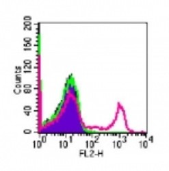
- Flow Cytometry: CD8 alpha Antibody (RPA-T8) [NBP2-25195] - Analysis using the PE conjugate of NBP2-25195. Staining of CD8 in 10^6 human PBMC using 10 ul (0.1 ug) of this antibody. The shaded histogram represents human PBMC alone; green represents mouse isotype control, red represents human CD8 this antibody.

- Submitted by
- Novus Biologicals (provider)
- Main image
- Experimental details
- Flow (Cell Surface): CD8 alpha Antibody (RPA-T8) [NBP2-25195] - Analysis using the PE conjugate of NBP2-25195. Staining of Human peripheral blood mononuclear cells. Image from verified customer review.
