 Explore
 Validate
 Learn
Learn Immunocytochemistry
 Immunohistochemistry
Antibody data
- Antibody Data
- Antigen structure
- References [4]
- Comments [0]
- Validations
- Immunocytochemistry [1]
Submit
Validation data
Reference
Comment
Report error
- Product number
- HPA001817 - Provider product page

- Provider
- Atlas Antibodies
- Proper citation
- Atlas Antibodies Cat#HPA001817, RRID:AB_1078779
- Product name
- Anti-CFB
- Antibody type
- Polyclonal
- Description
- Polyclonal Antibody against Human CFB, Gene description: complement factor B, Alternative Gene Names: BF, BFD, H2-Bf, Validated applications: IHC, ICC, Uniprot ID: P00751, Storage: Store at +4°C for short term storage. Long time storage is recommended at -20°C.
- Reactivity
- Human
- Host
- Rabbit
- Conjugate
- Unconjugated
- Isotype
- IgG
- Vial size
- 100 µl
- Concentration
- 0.1 mg/ml
- Storage
- Store at +4°C for short term storage. Long time storage is recommended at -20°C.
- Handling
- The antibody solution should be gently mixed before use.
Submitted references Insights into the Effects of Complement Factor H on the Assembly and Decay of the Alternative Pathway C3 Proconvertase and C3 Convertase
Variance decomposition of protein profiles from antibody arrays using a longitudinal twin model
Cancer genetics-guided discovery of serum biomarker signatures for diagnosis and prognosis of prostate cancer
Ca2+/calmodulin-dependent kinase II triggers cell membrane injury by inducing complement factor B gene expression in the mouse heart
Bettoni S, Bresin E, Remuzzi G, Noris M, Donadelli R
Journal of Biological Chemistry 2016;291(15):8214-8230a
Journal of Biological Chemistry 2016;291(15):8214-8230a
Variance decomposition of protein profiles from antibody arrays using a longitudinal twin model
Kato B, Nicholson G, Neiman M, Rantalainen M, Holmes C, Barrett A, Uhlén M, Nilsson P, Spector T, Schwenk J
Proteome Science 2011;9(1):73
Proteome Science 2011;9(1):73
Cancer genetics-guided discovery of serum biomarker signatures for diagnosis and prognosis of prostate cancer
Cima I, Schiess R, Wild P, Kaelin M, Schüffler P, Lange V, Picotti P, Ossola R, Templeton A, Schubert O, Fuchs T, Leippold T, Wyler S, Zehetner J, Jochum W, Buhmann J, Cerny T, Moch H, Gillessen S, Aebersold R, Krek W
Proceedings of the National Academy of Sciences 2011;108(8):3342-3347
Proceedings of the National Academy of Sciences 2011;108(8):3342-3347
Ca2+/calmodulin-dependent kinase II triggers cell membrane injury by inducing complement factor B gene expression in the mouse heart
Singh M, Kapoun A, Higgins L, Kutschke W, Thurman J, Zhang R, Singh M, Yang J, Guan X, Lowe J, Weiss R, Zimmermann K, Yull F, Blackwell T, Mohler P, Anderson M
Journal of Clinical Investigation 2009
Journal of Clinical Investigation 2009
No comments: Submit comment
Supportive validation
- Submitted by
- Atlas Antibodies (provider)
- Main image
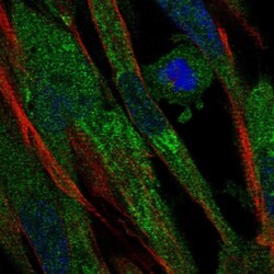
- Experimental details
- Immunofluorescent staining of human cell line ASC TERT1 shows localization to endoplasmic reticulum.
- Sample type
- Human
