 Explore
 Validate
 Learn
Learn Immunocytochemistry
Antibody data
- Antibody Data
- Antigen structure
- References [4]
- Comments [0]
- Validations
- Immunocytochemistry [1]
- Flow cytometry [1]
Submit
Validation data
Reference
Comment
Report error
- Product number
- MAB1249 - Provider product page

- Provider
- R&D Systems
- Product name
- Human/Mouse Glucagon Antibody
- Antibody type
- Monoclonal
- Description
- Protein A or G purified from hybridoma culture supernatant. Detects human and mouse Glucagon.
- Reactivity
- Human, Mouse
- Host
- Mouse
- Conjugate
- Unconjugated
- Isotype
- IgG
- Antibody clone number
- 181402
- Vial size
- 100 ug
- Concentration
- LYOPH
- Storage
- Use a manual defrost freezer and avoid repeated freeze-thaw cycles. 12 months from date of receipt, -20 to -70 °C as supplied. 1 month, 2 to 8 °C under sterile conditions after reconstitution. 6 months, -20 to -70 °C under sterile conditions after reconstitution.
Submitted references Abnormal neutrophil signature in the blood and pancreas of presymptomatic and symptomatic type 1 diabetes.
Neurogenin 3 Expressing Cells in the Human Exocrine Pancreas Have the Capacity for Endocrine Cell Fate.
Pancreatic islet cell phenotype and endocrine function throughout diabetes development in non-obese diabetic mice.
Stem cells derived from human fetal membranes display multilineage differentiation potential.
Vecchio F, Lo Buono N, Stabilini A, Nigi L, Dufort MJ, Geyer S, Rancoita PM, Cugnata F, Mandelli A, Valle A, Leete P, Mancarella F, Linsley PS, Krogvold L, Herold KC, Elding Larsson H, Richardson SJ, Morgan NG, Dahl-Jørgensen K, Sebastiani G, Dotta F, Bosi E, DRI_Biorepository Group, Type 1 Diabetes TrialNet Study Group, Battaglia M
JCI insight 2018 Sep 20;3(18)
JCI insight 2018 Sep 20;3(18)
Neurogenin 3 Expressing Cells in the Human Exocrine Pancreas Have the Capacity for Endocrine Cell Fate.
Gomez DL, O'Driscoll M, Sheets TP, Hruban RH, Oberholzer J, McGarrigle JJ, Shamblott MJ
PloS one 2015;10(8):e0133862
PloS one 2015;10(8):e0133862
Pancreatic islet cell phenotype and endocrine function throughout diabetes development in non-obese diabetic mice.
Kornete M, Beauchemin H, Polychronakos C, Piccirillo CA
Autoimmunity 2013 Jun;46(4):259-68
Autoimmunity 2013 Jun;46(4):259-68
Stem cells derived from human fetal membranes display multilineage differentiation potential.
Ilancheran S, Michalska A, Peh G, Wallace EM, Pera M, Manuelpillai U
Biology of reproduction 2007 Sep;77(3):577-88
Biology of reproduction 2007 Sep;77(3):577-88
No comments: Submit comment
Supportive validation
- Submitted by
- R&D Systems (provider)
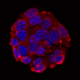
- Main image
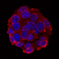
- Experimental details
- Glucagon in beta TC-6 Mouse Cell Line. Glucagon was detected in immersion fixed beta TC-6 mouse beta cell insulinoma cell line using 10 µg/mL Human/Mouse Glucagon Monoclonal Antibody (Catalog # MAB1249) for 3 hours at room temperature. Cells were stained with the NorthernLights™ 557-conjugated Anti-Mouse IgG Secondary Antibody (red; Catalog # NL007) and counterstained with DAPI (blue). View our protocol for Fluorescent ICC Staining of Cells on Coverslips.
Supportive validation
- Submitted by
- R&D Systems (provider)
- Main image
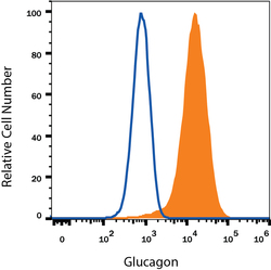
- Experimental details
- Detection of Glucagon in beta TC-6 Mouse Cell Line by Flow Cytometry. beta TC-6 mouse beta cell insulinoma cell line was stained with Mouse Anti-Human/Mouse Glucagon Monoclonal Antibody (Catalog # MAB1249, filled histogram) or isotype control antibody (Catalog # MAB003, open histogram), followed by Allophycocyanin-conjugated Anti-Mouse IgG Secondary Antibody (Catalog # F0101B). To facilitate intracellular staining, cells were fixed with Flow Cytometry Fixation Buffer (Catalog # FC004) and permeabilized with Flow Cytometry Permeabilization/Wash Buffer I (Catalog # FC005). View our protocol for Staining Intracellular Molecules.
