Explore
 Validate
 Learn
Learn Western blot
 ELISA
ELISAAntibody data
- Antibody Data
- Antigen structure
- References [1]
- Comments [0]
- Validations
- Western blot [3]
- Immunocytochemistry [6]
- Immunohistochemistry [5]
- Other assay [1]
Submit
Validation data
Reference
Comment
Report error
- Product number
- PA5-87938 - Provider product page

- Provider
- Invitrogen Antibodies
- Product name
- MTAP Polyclonal Antibody
- Antibody type
- Polyclonal
- Antigen
- Recombinant full-length protein
- Description
- Immunogen sequence: MASGTTTTAV KIGIIGGTGL DDPEILEGRT EKYVDTPFGK PSDALILGKI KNVDCVLLAR HGRQHTIMPS KVNYQANIWA LKEEGCTHVI VTTACGSLRE EIQPGDIVII DQFIDRTTMR PQSFYDGSHS CARGVCHIPM AEPFCPKTRE VLIETAKKLG LRCHSKGTMV TIEGPRFSSR AESFMFRTWG ADVINMTTVP EVVLAKEAGI CYASIAMATD YDCWKEHEEA VSVDRVLKTL KENANKAKSL LLTTIPQIGS TEWSETLHNL KNMAQFSVLL PRH; Positive Samples: 22Rv1, 293T, Mouse liver; Cellular Location: Cytoplasm, Nucleus
- Reactivity
- Human, Mouse, Rat
- Host
- Rabbit
- Isotype
- IgG
- Vial size
- 100 μL
- Concentration
- 0.6 mg/mL
- Storage
- -20°C, Avoid Freeze/Thaw Cycles
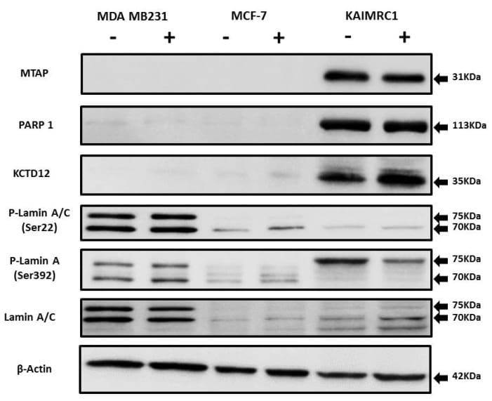
Submitted references Proteomics Profiling of KAIMRC1 in Comparison to MDA-MB231 and MCF-7.
Alghanem B, Ali R, Nehdi A, Al Zahrani H, Altolayyan A, Shaibah H, Baz O, Alhallaj A, Moresco JJ, Diedrich JK, Yates JR 3rd, Boudjelal M
International journal of molecular sciences 2020 Jun 18;21(12)
International journal of molecular sciences 2020 Jun 18;21(12)
No comments: Submit comment
Supportive validation
- Submitted by
- Invitrogen Antibodies (provider)
- Main image

- Experimental details
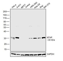
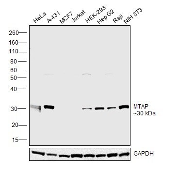
- Western blot analysis of MTAP in extracts of various cell lines. Samples were incubated with MTAP Polyclonal antibody (Product # PA5-87938) using a dilution of 1:1,000, followed by HRP Goat Anti-Rabbit IgG (H+L) at a dilution of 1:10,000. Lysates/proteins: 25 µg per lane. Blocking buffer: 3% nonfat dry milk in TBST. Detection: ECL Basic Kit. Exposure time: 180s.
- Submitted by
- Invitrogen Antibodies (provider)
- Main image
- Experimental details
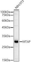
- Western blot analysis of MTAP in extracts of NIH/3T3 cells. Samples were incubated with MTAP Polyclonal antibody (Product # PA5-87938) using a dilution of 1:1,000, followed by HRP Goat Anti-Rabbit IgG (H+L) at a dilution of 1:10,000. Lysates/proteins: 25 µg per lane. Blocking buffer: 3% nonfat dry milk in TBST. Detection: ECL Basic Kit. Exposure time: 30s.

- Submitted by
- Invitrogen Antibodies (provider)
- Main image
- Experimental details
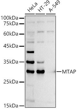
- Western blot was performed using Anti-MTAP Polyclonal Antibody (Product # PA5-87938) and a 32 kDa band corresponding to S-methyl-5-thioadenosine phosphorylase was observed across all positive cell lines tested, and not in negative cell lines (MCF7 and Jurkat). Whole cell extracts (30 µg lysate) of HeLa (Lane 1), A-431 (Lane 2), MCF7 (Lane 3), Jurkat (Lane 4), HEK-293 (Lane 5), HepG2 (Lane 6), Raji (Lane 7) and NIH 3T3 (Lane 8) were electrophoresed using NuPAGE™ 12% Bis-Tris Protein Gel (Product # NP0341BOX). Resolved proteins were then transferred onto a Nitrocellulose membrane (Product # IB23001) by iBlot® 2 Dry Blotting System (Product # IB21001). The blot was probed with the primary antibody (1:1000 dilution) and detected by chemiluminescence with Goat anti-Rabbit IgG (Heavy Chain) Superclonal™ Recombinant Secondary Antibody, HRP (Product # A27036, 1:4000 dilution) using the iBright FL 1000 (Product # A32752). Chemiluminescent detection was performed using Novex® ECL Reagent Kit (Product # WP20005).
Supportive validation
- Submitted by
- Invitrogen Antibodies (provider)
- Main image
- Experimental details
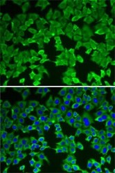
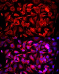
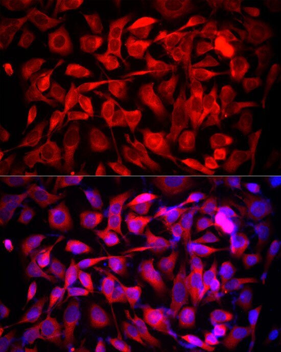
- Immunocytochemistry-Immunofluorescence analysis of MTAP was performed in A-549 cells using MTAP Polyclonal Antibody (Product # PA5-87938).
- Submitted by
- Invitrogen Antibodies (provider)
- Main image
- Experimental details
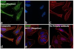
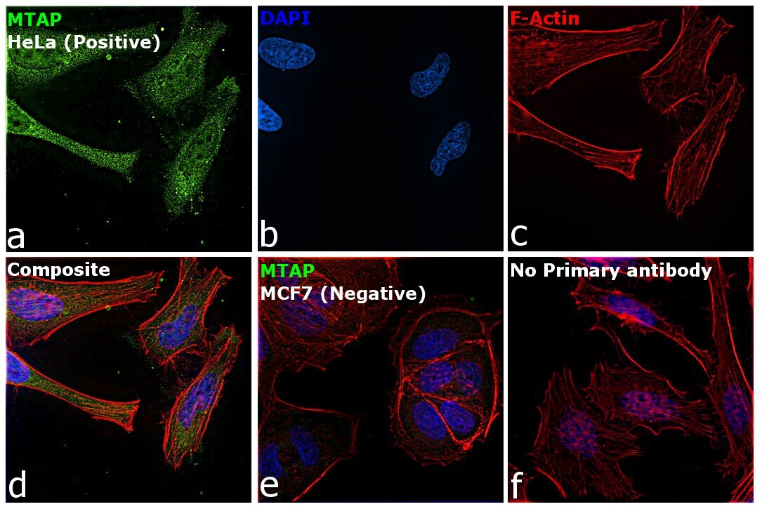
- Immunofluorescence analysis of S-methyl-5-thioadenosine phosphorylase was performed using 70% confluent log phase HeLa and MCF7 cell lines. The cells were fixed with 4% paraformaldehyde for 10 minutes, permeabilized with 0.1% Triton™ X-100 for 15 minutes, and blocked with 2% BSA for 45 minutes at room temperature. The cells were labeled with MTAP Polyclonal Antibody (Product # PA5-87938) at 1:100 dilution in 0.1% BSA, incubated at 4 degree celsius overnight and then labeled with Donkey anti-Rabbit IgG (H+L) Highly Cross-Adsorbed Secondary Antibody, Alexa Fluor Plus 488 (Product # A32790), (1:2000 dilution), for 45 minutes at room temperature (Panel a: Green). Nuclei (Panel b: Blue) were stained with ProLong™ Diamond Antifade Mountant with DAPI (Product # P36962). F-actin (Panel c: Red) was stained with Rhodamine Phalloidin (Product # R415, 1:300 dilution). Panel d represents the merged image showing cytoplasmic and nuclear localization. Panel e represents MCF7 cell line which does not express MTAP protein. Panel f represents control cells with no primary antibody to assess background. The images were captured at 60X magnification.

- Submitted by
- Invitrogen Antibodies (provider)
- Main image
- Experimental details
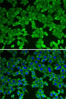
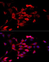
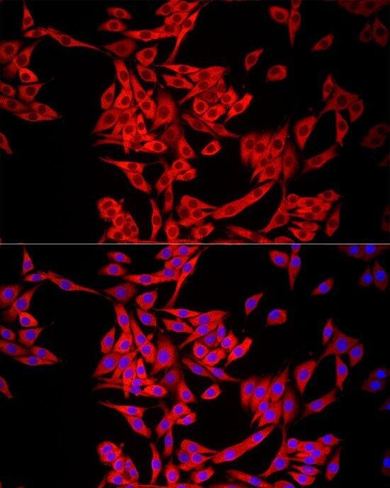
- Immunofluorescence analysis of MTAP in NIH/3T3 cells. Samples were incubated with MTAP Polyclonal antibody (Product # PA5-87938) using a dilution of 1:50 (40x lens). Blue: DAPI for nuclear staining.

- Submitted by
- Invitrogen Antibodies (provider)
- Main image
- Experimental details
- Immunofluorescence analysis of MTAP in PC-12 cells. Samples were incubated with MTAP Polyclonal antibody (Product # PA5-87938) using a dilution of 1:50 (40x lens). Blue: DAPI for nuclear staining.
- Submitted by
- Invitrogen Antibodies (provider)
- Main image
- Experimental details
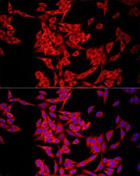
- Immunofluorescence analysis of MTAP in HeLa cells. Samples were incubated with MTAP Polyclonal antibody (Product # PA5-87938) using a dilution of 1:50 (40x lens). Blue: DAPI for nuclear staining.
- Submitted by
- Invitrogen Antibodies (provider)
- Main image

- Experimental details
- Immunofluorescence analysis of S-methyl-5-thioadenosine phosphorylase was performed using 70% confluent log phase HeLa and MCF7 cell lines. The cells were fixed with 4% paraformaldehyde for 10 minutes, permeabilized with 0.1% Triton™ X-100 for 15 minutes, and blocked with 2% BSA for 45 minutes at room temperature. The cells were labeled with MTAP Polyclonal Antibody (Product # PA5-87938) at 1:100 dilution in 0.1% BSA, incubated at 4 degree celsius overnight and then labeled with Donkey anti-Rabbit IgG (H+L) Highly Cross-Adsorbed Secondary Antibody, Alexa Fluor Plus 488 (Product # A32790), (1:2000 dilution), for 45 minutes at room temperature (Panel a: Green). Nuclei (Panel b: Blue) were stained with ProLong™ Diamond Antifade Mountant with DAPI (Product # P36962). F-actin (Panel c: Red) was stained with Rhodamine Phalloidin (Product # R415, 1:300 dilution). Panel d represents the merged image showing cytoplasmic and nuclear localization. Panel e represents MCF7 cell line which does not express MTAP protein. Panel f represents control cells with no primary antibody to assess background. The images were captured at 60X magnification.
Supportive validation
- Submitted by
- Invitrogen Antibodies (provider)
- Main image

- Experimental details
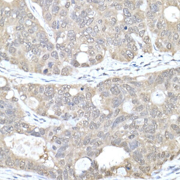
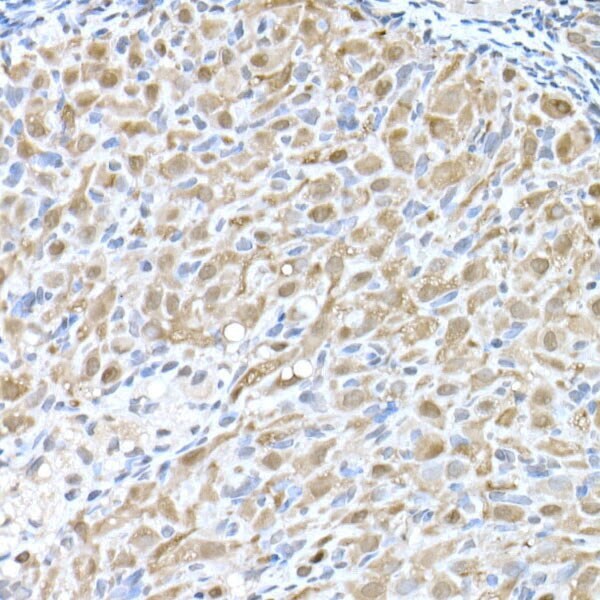
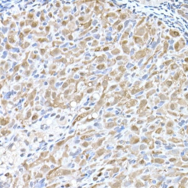
- Immunohistochemistry analysis of MTAP in paraffin-embedded human colon carcinoma. Samples were incubated with MTAP Polyclonal antibody (Product # PA5-87938) using a dilution of 1:50 (40x lens). Perform high pressure antigen retrieval with 10 mM citrate buffer pH 6.0 before commencing with IHC staining protocol.
- Submitted by
- Invitrogen Antibodies (provider)
- Main image

- Experimental details
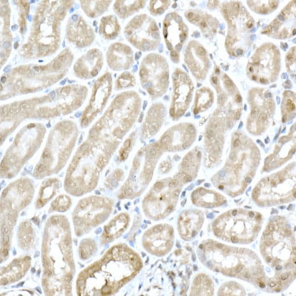
- Immunohistochemistry analysis of MTAP in paraffin-embedded mouse kidney. Samples were incubated with MTAP Polyclonal antibody (Product # PA5-87938) using a dilution of 1:50 (40x lens). Perform high pressure antigen retrieval with 10 mM citrate buffer pH 6.0 before commencing with IHC staining protocol.
- Submitted by
- Invitrogen Antibodies (provider)
- Main image

- Experimental details
- Immunohistochemistry analysis of MTAP in paraffin-embedded rat ovary. Samples were incubated with MTAP Polyclonal antibody (Product # PA5-87938) using a dilution of 1:50 (40x lens). Perform high pressure antigen retrieval with 10 mM citrate buffer pH 6.0 before commencing with IHC staining protocol.
- Submitted by
- Invitrogen Antibodies (provider)
- Main image
- Experimental details
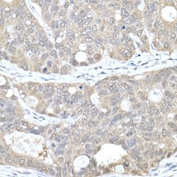
- Immunohistochemistry analysis of MTAP in paraffin-embedded human colon carcinoma. Samples were incubated with MTAP Polyclonal antibody (Product # PA5-87938) using a dilution of 1:50 (40x lens). Perform high pressure antigen retrieval with 10 mM citrate buffer pH 6.0 before commencing with IHC staining protocol.

- Submitted by
- Invitrogen Antibodies (provider)
- Main image

- Experimental details
- Immunohistochemistry analysis of MTAP in paraffin-embedded rat ovary. Samples were incubated with MTAP Polyclonal antibody (Product # PA5-87938) using a dilution of 1:50 (40x lens). Perform high pressure antigen retrieval with 10 mM citrate buffer pH 6.0 before commencing with IHC staining protocol.
Supportive validation
- Submitted by
- Invitrogen Antibodies (provider)
- Main image
- Experimental details
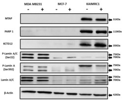
- Figure 5 Expression of MTAP, PARP1, KCTD12, and Lamin A/C in cancer cell lines: Western blot analysis of protein expression of MTAP, PARP1, KCTD12, Lamin A/C, and phosphorylated Lamin A/C in MDA MB231, MCF-7, and KAIMRC1 cells. Preparation of cell lysates and western blot were performed as described under the Materials and Methods section. In both normal (+) and serum-starved (-) conditions, KAIMRC1 cells showed strong expression of MTAP, PARP1, and KCTD12 compared to MDA-MB231 and MCF-7 cells. Moreover, KAIMRC1 cells showed weak expression of Lamin A/C and phosphorylated Lamin A/C in comparison to MDA MB231 cells, thus validating our proteomics results.