Explore
 Validate
 Learn
Learn Western blot
 Flow cytometry
Antibody data
- Antibody Data
- Antigen structure
- References [2]
- Comments [0]
- Validations
- Western blot [1]
Submit
Validation data
Reference
Comment
Report error
- Product number
- PB9103 - Provider product page

- Provider
- Boster Biological Technology
- Product name
- Anti-Cyclin D3/CCND3 Antibody Picoband™
- Antibody type
- Polyclonal
- Description
- Polyclonal antibody for Cyclin D3/CCND3 detection. Host: Rabbit.Size: 100μg/vial. Tested applications: WB. Reactive species: Human. Cyclin D3/CCND3 information: Molecular Weight: 32520 MW; Subcellular Localization: Nucleus . Cytoplasm . Membrane . Cyclin D-CDK4 complexes accumulate at the nuclear membrane and are then translocated to the nucleus through interaction with KIP/CIP family members.
- Reactivity
- Human, Rat
- Host
- Rabbit
- Vial size
- 100μg/vial
- Concentration
- Add 0.2ml of distilled water will yield a concentration of 500ug/ml.
- Storage
- At -20°C for one year. After reconstitution, at 4°C for one month. It can also be aliquoted and stored frozen at -20°C for a longer time. Avoid repeated freezing and thawing.
- Handling
- Add 0.2ml of distilled water will yield a concentration of 500ug/ml.
Submitted references Co-culture with TM4 cells enhances the proliferation and migration of rat adipose-derived mesenchymal stem cells with high stemness.
Wall shear stress promotes intimal hyperplasia through the paracrine H(2)O(2)-mediated NOX-AKT-SVV axis.
Luo Y, Mohsin A, Xu C, Wang Q, Hang H, Zhuang Y, Chu J, Guo M
Cytotechnology 2018 Oct;70(5):1409-1422
Cytotechnology 2018 Oct;70(5):1409-1422
Wall shear stress promotes intimal hyperplasia through the paracrine H(2)O(2)-mediated NOX-AKT-SVV axis.
Zhang H, Yang Z, Wang J, Wang X, Zhao Y, Zhu F
Life sciences 2018 Aug 15;207:61-71
Life sciences 2018 Aug 15;207:61-71
No comments: Submit comment
Supportive validation
- Submitted by
- Boster Biological Technology (provider)
- Main image

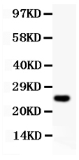
- Experimental details
- Western blot analysis of Cyclin D3 using anti-Cyclin D3 antibody (PB9103). Electrophoresis was performed on a 5-20% SDS-PAGE gel at 70V (Stacking gel) / 90V (Resolving gel) for 2-3 hours. lane 1: recombinant human Cyclin D3 protein 0.5ng. After Electrophoresis, proteins were transferred to a Nitrocellulose membrane at 150mA for 50-90 minutes. Blocked the membrane with 5% Non-fat Milk/ TBS for 1.5 hour at RT. The membrane was incubated with rabbit anti-Cyclin D3 antigen affinity purified polyclonal antibody (Catalog # PB9103) at 0.5 μg/mL overnight at 4°C, then washed with TBS-0.1%Tween 3 times with 5 minutes each and probed with a goat anti-rabbit IgG-HRP secondary antibody at a dilution of 1:10000 for 1.5 hour at RT. The signal is developed using an Enhanced Chemiluminescent detection (ECL) kit (Catalog # EK1002) with Tanon 5200 system. A specific band was detected for Cyclin D3 at approximately 25KD. The expected band size for Cyclin D3 is at 25KD.
- Additional image