Explore
 Validate
 Learn
Learn Western blot
Antibody data
- Antibody Data
- Antigen structure
- References [6]
- Comments [0]
- Validations
- Western blot [2]
- Immunohistochemistry [3]
- Other assay [1]
Submit
Validation data
Reference
Comment
Report error
- Product number
- MA5-12726 - Provider product page

- Provider
- Invitrogen Antibodies
- Product name
- Anti-Cyclin D3 Monoclonal Antibody (DCS-28.1)
- Antibody type
- Monoclonal
- Antigen
- Recombinant full-length protein
- Description
- MA5-12726 targets Cyclin D3 in WB applications and shows reactivity with Human and Rat samples. The MA5-12726 immunogen is purified human recombinant full length cyclin D3 protein.
- Reactivity
- Human, Mouse, Rat
- Host
- Mouse
- Isotype
- IgG
- Antibody clone number
- DCS-28.1
- Vial size
- 500 µL
- Concentration
- 0.2 mg/mL
- Storage
- 4° C
Submitted references CDK4 T172 phosphorylation is central in a CDK7-dependent bidirectional CDK4/CDK2 interplay mediated by p21 phosphorylation at the restriction point.
Combined effect of cyclin D3 expression and abrogation of cyclin D1 prevent mouse skin tumor development.
Differential utilization of cyclin D1 and cyclin D3 in the distinct mitogenic stimulations by growth factors and TSH of human thyrocytes in primary culture.
Differential involvement of the actin cytoskeleton in differentiation and mitogenesis of thyroid cells: inactivation of Rho proteins contributes to cyclic adenosine monophosphate-dependent gene expression but prevents mitogenesis.
Targeted disruption of CDK4 delays cell cycle entry with enhanced p27(Kip1) activity.
Targeted disruption of CDK4 delays cell cycle entry with enhanced p27(Kip1) activity.
Bisteau X, Paternot S, Colleoni B, Ecker K, Coulonval K, De Groote P, Declercq W, Hengst L, Roger PP
PLoS genetics 2013 May;9(5):e1003546
PLoS genetics 2013 May;9(5):e1003546
Combined effect of cyclin D3 expression and abrogation of cyclin D1 prevent mouse skin tumor development.
Wang X, Sistrunk C, Miliani de Marval PL, Kim Y, Rodriguez-Puebla ML
Cell cycle (Georgetown, Tex.) 2012 Jan 15;11(2):335-42
Cell cycle (Georgetown, Tex.) 2012 Jan 15;11(2):335-42
Differential utilization of cyclin D1 and cyclin D3 in the distinct mitogenic stimulations by growth factors and TSH of human thyrocytes in primary culture.
Paternot S, Dumont JE, Roger PP
Molecular endocrinology (Baltimore, Md.) 2006 Dec;20(12):3279-92
Molecular endocrinology (Baltimore, Md.) 2006 Dec;20(12):3279-92
Differential involvement of the actin cytoskeleton in differentiation and mitogenesis of thyroid cells: inactivation of Rho proteins contributes to cyclic adenosine monophosphate-dependent gene expression but prevents mitogenesis.
Fortemaison N, Blancquaert S, Dumont JE, Maenhaut C, Aktories K, Roger PP, Dremier S
Endocrinology 2005 Dec;146(12):5485-95
Endocrinology 2005 Dec;146(12):5485-95
Targeted disruption of CDK4 delays cell cycle entry with enhanced p27(Kip1) activity.
Tsutsui T, Hesabi B, Moons DS, Pandolfi PP, Hansel KS, Koff A, Kiyokawa H
Molecular and cellular biology 1999 Oct;19(10):7011-9
Molecular and cellular biology 1999 Oct;19(10):7011-9
Targeted disruption of CDK4 delays cell cycle entry with enhanced p27(Kip1) activity.
Tsutsui T, Hesabi B, Moons DS, Pandolfi PP, Hansel KS, Koff A, Kiyokawa H
Molecular and cellular biology 1999 Oct;19(10):7011-9
Molecular and cellular biology 1999 Oct;19(10):7011-9
No comments: Submit comment
Supportive validation
- Submitted by
- Invitrogen Antibodies (provider)
- Main image

- Experimental details
- Western blot of Cyclin D3 using Cyclin D3 Monoclonal Antibody (Product # MA5-12726) on PC12 Cells.
- Submitted by
- Invitrogen Antibodies (provider)
- Main image
- Experimental details
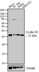
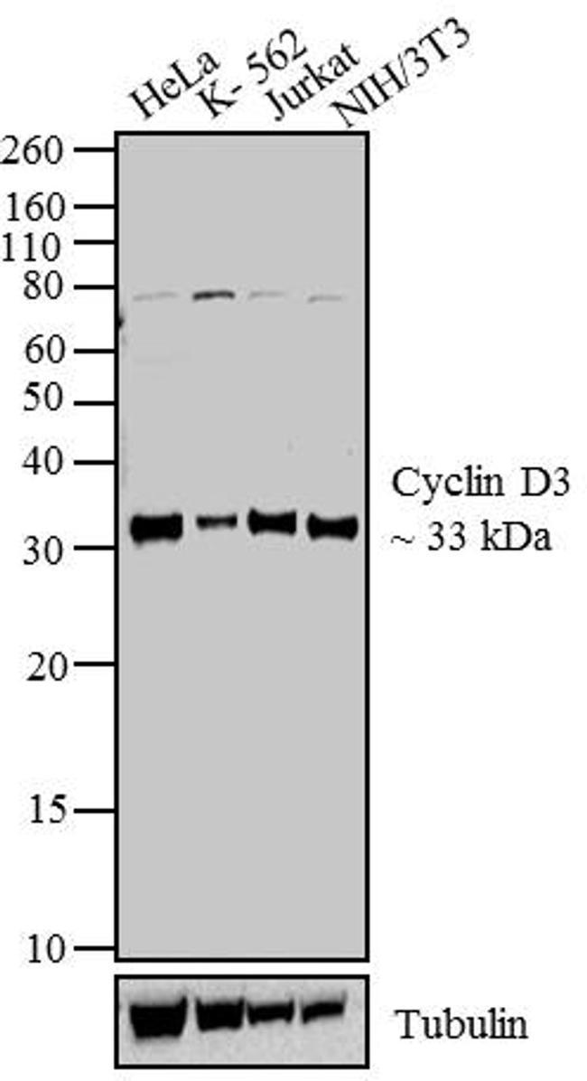
- Western blot analysis was performed using nuclear enriched extracts (30 µg) of HeLa (Lane 1), K-562 (Lane 2), Jurkat (Lane 3) and NIH/3T3 (Lane 4). The blots was probed with Anti-Cyclin D3 Mouse Monoclonal Antibody (Product # MA5-12726, 2 µg/mL) and detected by chemiluminescence using Goat anti-Mouse IgG (H+L) Superclonal™ Secondary Antibody, HRP conjugate (Product # A28177, 0.4 µg/mL, 1:2500 dilution). A ~ 33 kDa band corresponding to Cyclin D3 was observed across cell lines tested. Known quantity of protein samples were electrophoresed using Novex® NuPAGE®12 % Bis-Tris gel (Product # NP0342BOX), XCell SureLock™ Electrophoresis System (Product # EI0002) and Novex® Sharp Pre-Stained Protein Standard (Product # LC5800). Resolved proteins were then transferred onto a nitrocellulose membrane with iBlot® 2 Dry Blotting System (Product # IB21001). The membrane was probed with the relevant primary and secondary Antibody using iBind™ Flex Western Starter Kit (Product # SLF2000S). Chemiluminescent detection was performed using Pierce™ ECL Western Blotting Substrate (Product # 32106).
Supportive validation
- Submitted by
- Invitrogen Antibodies (provider)
- Main image

- Experimental details
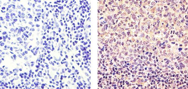
- Immunohistochemistry analysis of Cyclin D3 showing staining in the nucleus of paraffin-embedded human tonsil tissue (right) compared to a negative control without primary antibody (left). To expose target proteins, antigen retrieval was performed using 10mM sodium citrate (pH 6.0), microwaved for 8-15 min. Following antigen retrieval, tissues were blocked in 3% H2O2-methanol for 15 min at room temperature, washed with ddH2O and PBS, and then probed with a Cyclin D3 Antibody Mouse Monoclonal Antibody (Product # MA5-12726) diluted in 3% BSA-PBS at a dilution of 1:20 for 1 hour at 37°C in a humidified chamber. Tissues were washed extensively in PBST and detection was performed using an HRP-conjugated secondary antibody followed by colorimetric detection using a DAB kit. Tissues were counterstained with hematoxylin and dehydrated with ethanol and xylene to prep for mounting.
- Submitted by
- Invitrogen Antibodies (provider)
- Main image

- Experimental details
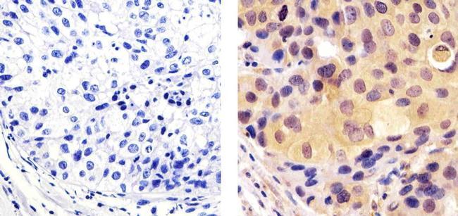
- Immunohistochemistry analysis of Cyclin D3 showing staining in the nucleus of paraffin-embedded human breast carcinoma (right) compared to a negative control without primary antibody (left). To expose target proteins, antigen retrieval was performed using 10mM sodium citrate (pH 6.0), microwaved for 8-15 min. Following antigen retrieval, tissues were blocked in 3% H2O2-methanol for 15 min at room temperature, washed with ddH2O and PBS, and then probed with a Cyclin D3 Antibody Mouse Monoclonal Antibody (Product # MA5-12726) diluted in 3% BSA-PBS at a dilution of 1:50 for 1 hour at 37°C in a humidified chamber. Tissues were washed extensively in PBST and detection was performed using an HRP-conjugated secondary antibody followed by colorimetric detection using a DAB kit. Tissues were counterstained with hematoxylin and dehydrated with ethanol and xylene to prep for mounting.
- Submitted by
- Invitrogen Antibodies (provider)
- Main image
- Experimental details
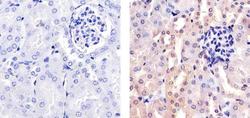
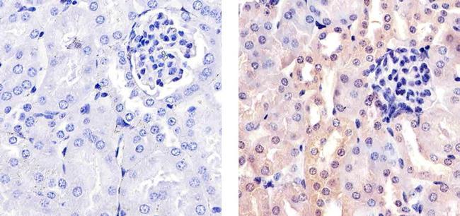
- Immunohistochemistry analysis of Cyclin D3 showing staining in the nucleus of paraffin-embedded mouse kidney tissue (right) compared to a negative control without primary antibody (left). To expose target proteins, antigen retrieval was performed using 10mM sodium citrate (pH 6.0), microwaved for 8-15 min. Following antigen retrieval, tissues were blocked in 3% H2O2-methanol for 15 min at room temperature, washed with ddH2O and PBS, and then probed with a Cyclin D3 Antibody Mouse Monoclonal Antibody (Product # MA5-12726) diluted in 3% BSA-PBS at a dilution of 1:20 for 1 hour at 37°C in a humidified chamber. Tissues were washed extensively in PBST and detection was performed using an HRP-conjugated secondary antibody followed by colorimetric detection using a DAB kit. Tissues were counterstained with hematoxylin and dehydrated with ethanol and xylene to prep for mounting.
Supportive validation
- Submitted by
- Invitrogen Antibodies (provider)
- Main image

- Experimental details
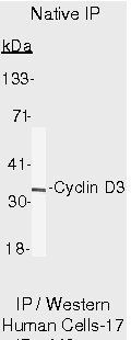
- Immunoprecipitation of Cyclin D3 using Cyclin D3 Monoclonal Antibody (Product # MA5-12726) on Native Human LS174T Cells.