 Explore
 Validate
 Learn
Learn Western blot
Antibody data
- Antibody Data
- Antigen structure
- References [3]
- Comments [0]
- Validations
- Western blot [1]
- Flow cytometry [1]
Submit
Validation data
Reference
Comment
Report error
- Product number
- AF5729 - Provider product page

- Provider
- Novus Biologicals
- Product name
- Goat Polyclonal WT1 Antibody
- Antibody type
- Polyclonal
- Description
- Antigen Affinity-purified. Detects human WT1 in direct ELISAs and Western blots.
- Reactivity
- Human
- Host
- Goat
- Conjugate
- Unconjugated
- Isotype
- IgG
- Vial size
- 100 ug
- Concentration
- LYOPH
- Storage
- Use a manual defrost freezer and avoid repeated freeze-thaw cycles. 12 months from date of receipt, -20 to -70 degreesC as supplied. 1 month, 2 to 8 degreesC under sterile conditions after reconstitution. 6 months, -20 to -70 degreesC under sterile conditions after reconstitution.
Submitted references Differentiation of human iPSCs into functional podocytes.
Renal progenitors derived from human iPSCs engraft and restore function in a mouse model of acute kidney injury.
A novel source of cultured podocytes.
Rauch C, Feifel E, Kern G, Murphy C, Meier F, Parson W, Beilmann M, Jennings P, Gstraunthaler G, Wilmes A
PloS one 2018;13(9):e0203869
PloS one 2018;13(9):e0203869
Renal progenitors derived from human iPSCs engraft and restore function in a mouse model of acute kidney injury.
Imberti B, Tomasoni S, Ciampi O, Pezzotta A, Derosas M, Xinaris C, Rizzo P, Papadimou E, Novelli R, Benigni A, Remuzzi G, Morigi M
Scientific reports 2015 Mar 6;5:8826
Scientific reports 2015 Mar 6;5:8826
A novel source of cultured podocytes.
Da Sacco S, Lemley KV, Sedrakyan S, Zanusso I, Petrosyan A, Peti-Peterdi J, Burford J, De Filippo RE, Perin L
PloS one 2013;8(12):e81812
PloS one 2013;8(12):e81812
No comments: Submit comment
Supportive validation
- Submitted by
- Novus Biologicals (provider)
- Main image
- Experimental details
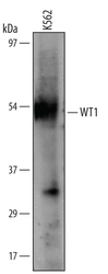
- Detection of Human WT1 by Western Blot. Western blot shows lysates of K562 human chronic myelogenous leukemia cell line. PVDF membrane was probed with 1 µg/mL of Goat Anti-Human WT1 Antigen Affinity-purified Polyclonal Antibody (Catalog # AF5729) followed by HRP-conjugated Anti-Goat IgG Secondary Antibody (Catalog # HAF019). A specific band was detected for WT1 at approximately 54 kDa (as indicated). This experiment was conducted under reducing conditions and using Immunoblot Buffer Group 8.

Supportive validation
- Submitted by
- Novus Biologicals (provider)
- Main image

- Experimental details
- Detection of WT1 in HL-60 Human Cell Line by Flow Cytometry. HL-60 human acute promyelocytic leukemia cell line was stained with Goat Anti-Human WT1 Antigen Affinity-purified Polyclonal Antibody (Catalog # AF5729, filled histogram) or control antibody (Catalog # AB-108-C, open histogram), followed by Allophycocyanin-conjugated Anti-Goat IgG Secondary Antibody (Catalog # F0108). To facilitate intracellular staining, cells were fixed with paraformaldehyde and permeabilized with saponin.
