 Explore
 Validate
 Learn
Learn Western blot
Antibody data
- Antibody Data
- Antigen structure
- References [4]
- Comments [0]
- Validations
- Western blot [1]
- Immunocytochemistry [1]
- Immunohistochemistry [1]
Submit
Validation data
Reference
Comment
Report error
- Product number
- ABIN955586 - Provider product page

- Provider
- antibodies-online
- Product name
- anti-Wilms Tumor 1 (WT1) (AA 353-383), (Middle Region) antibody
- Antibody type
- Polyclonal
- Description
- Protein A column followed by peptide affinity purification
- Reactivity
- Human
- Host
- Rabbit
- Epitope
- AA 353-383, Middle Region
- Vial size
- 0.4 mL
- Storage
- Store at 2 - 8°C for up to six months or (in aliquots) at -20°C for longer.
- Handling
- Avoid repeated freezing and thawing.
Submitted references Wilms' tumour 1 can suppress hTERT gene expression and telomerase activity in clear cell renal cell carcinoma via multiple pathways.
WT1 expression correlates with angiogenesis in endometrial cancer tissue.
The complex life of WT1.
Paternal expression of WT1 in human fibroblasts and lymphocytes.
Sitaram RT, Degerman S, Ljungberg B, Andersson E, Oji Y, Sugiyama H, Roos G, Li A
British journal of cancer 2010 Oct 12;103(8):1255-62
British journal of cancer 2010 Oct 12;103(8):1255-62
WT1 expression correlates with angiogenesis in endometrial cancer tissue.
Dohi S, Ohno S, Ohno Y, Kyo S, Soma G, Sugiyama H, Inoue M
Anticancer research 2010 Aug;30(8):3187-92
Anticancer research 2010 Aug;30(8):3187-92
The complex life of WT1.
Wagner KD, Wagner N, Schedl A
Journal of cell science 2003 May 1;116(Pt 9):1653-8
Journal of cell science 2003 May 1;116(Pt 9):1653-8
Paternal expression of WT1 in human fibroblasts and lymphocytes.
Mitsuya K, Sui H, Meguro M, Kugoh H, Jinno Y, Niikawa N, Oshimura M
Human molecular genetics 1997 Dec;6(13):2243-6
Human molecular genetics 1997 Dec;6(13):2243-6
No comments: Submit comment
Supportive validation
- Submitted by
- antibodies-online (provider)
- Main image
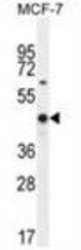
- Experimental details
- WB

Supportive validation
- Submitted by
- antibodies-online (provider)
- Main image
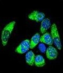
- Experimental details
- Confocal immunofluorescent analysis of WT1 Antibody (Center E361)(AP54572PU-N) with MCF-7 cell followed by Alexa Fluor® 488-conjugated goat anti-rabbit lgG (green). DAPI was used to stain the cell nuclear (blue).

Supportive validation
- Submitted by
- antibodies-online (provider)
- Main image

- Experimental details
- IHC
