Explore
 Validate
 Learn
Learn Western blot
 ELISA
ELISAAntibody data
- Antibody Data
- Antigen structure
- References [0]
- Comments [0]
- Validations
- Western blot [1]
- Immunocytochemistry [2]
- Immunohistochemistry [1]
- Flow cytometry [2]
Submit
Validation data
Reference
Comment
Report error
- Product number
- MA5-33113 - Provider product page

- Provider
- Invitrogen Antibodies
- Product name
- Actin Recombinant Rabbit Monoclonal Antibody (25E3)
- Antibody type
- Monoclonal
- Antigen
- Other
- Reactivity
- Human
- Host
- Rabbit
- Isotype
- IgG
- Antibody clone number
- 25E3
- Vial size
- 100 μL
- Concentration
- 0.4 mg/mL
- Storage
- -20°C or -80°C if preferred
No comments: Submit comment
Supportive validation
- Submitted by
- Invitrogen Antibodies (provider)
- Main image
- Experimental details
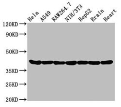
- Western Blot analysis of Actin using a Actin Monoclonal antibody (Product # MA5-33113) at a concentration of 0.95 µg/mL. Positive WB detected in: Hela whole cell lysate, A549 whole cell lysate, Raw264.7 whole cell lysate, NIH/3T3 whole cell lysate, HepG2 whole cell lysate, Rat brain tissue, Rat heart tissue. All lanes: Actin antibody at 0.95µg/ml. A secondary Goat polyclonal antibody to rabbit IgG was applied at a 1:50,000 dilution. Observed band size: 42 kDa.

Supportive validation
- Submitted by
- Invitrogen Antibodies (provider)
- Main image
- Experimental details
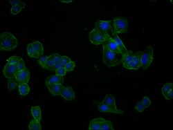
- Immunofluorescent analysis of Actin in HepG2 cells using a Actin monoclonal antibody (Product # MA5-33113) at a dilution of 1:60. The cells were fixed in 4% formaldehyde, permeabilized using 0.2% Triton X-100 and blocked in 10% normal Goat Serum. The cells were then incubated with the antibody overnight at 4°C.The secondary antibody was Alexa Fluor 488-congugated Goat Anti-Rabbit IgG (H+L). Cells were counter-stained with DAPI.

- Submitted by
- Invitrogen Antibodies (provider)
- Main image

- Experimental details
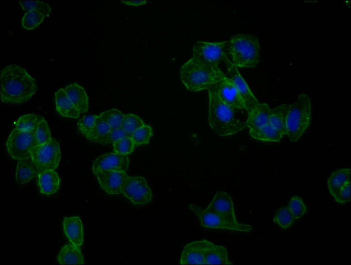
- Immunofluorescent analysis of Actin in HepG2 cells using a Actin monoclonal antibody (Product # MA5-33113) at a dilution of 1:60. The cells were fixed in 4% formaldehyde, permeabilized using 0.2% Triton X-100 and blocked in 10% normal Goat Serum. The cells were then incubated with the antibody overnight at 4°C.The secondary antibody was Alexa Fluor 488-congugated Goat Anti-Rabbit IgG (H+L). Cells were counter-stained with DAPI.
Supportive validation
- Submitted by
- Invitrogen Antibodies (provider)
- Main image
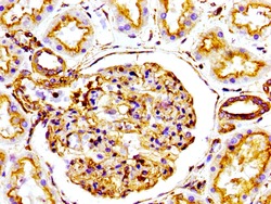
- Experimental details
- Immunohistochemical analysis of Actin in paraffin embedded human kidney tissue using a Actin monoclonal antibody (Product # MA5-33113) at a dilution of 1:100. After dewaxing and hydration, antigen retrieval was mediated by high pressure in a citrate buffer (pH 6.0). Section was blocked with 10% normal goat serum 30min at RT. Then primary antibody (1% BSA) was incubated at 4°C overnight. The primary is detected by a biotinylated secondary antibody and visualized using an HRP conjugated SP system.

Supportive validation
- Submitted by
- Invitrogen Antibodies (provider)
- Main image
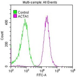
- Experimental details
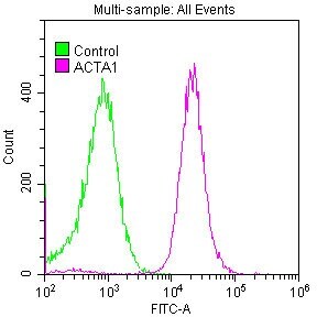
- Flow cytometric analysis of Actin in Hela cells using a monoclonal antibody (Product # MA5-33113) at a dilution of 1:50. The cells were fixed with 70% Ethyl alcohol (18h) and then permeabilized with 0.3% Triton X-100 for 2 min. The cells were then incubated in 1x PBS /10% normal goat serum to block non-specific protein-protein interactions followed by primary antibody for 1 h at 4°C. The secondary antibody used was FITC goat anti-rabbit IgG (H+L) at 1:200 dilution for 1 h at 4°C. Control antibody (green line) was used under the same conditions. Acquisition of >10,000 events was performed.

- Submitted by
- Invitrogen Antibodies (provider)
- Main image

- Experimental details
- Flow cytometric analysis of Actin in Hela cells using a monoclonal antibody (Product # MA5-33113) at a dilution of 1:50. The cells were fixed with 70% Ethyl alcohol (18h) and then permeabilized with 0.3% Triton X-100 for 2 min. The cells were then incubated in 1x PBS /10% normal goat serum to block non-specific protein-protein interactions followed by primary antibody for 1 h at 4°C. The secondary antibody used was FITC goat anti-rabbit IgG (H+L) at 1:200 dilution for 1 h at 4°C. Control antibody (green line) was used under the same conditions. Acquisition of >10,000 events was performed.