 Explore
 Validate
 Learn
Learn Western blot
 Immunoprecipitation
 Immunohistochemistry
Antibody data
- Antibody Data
- Antigen structure
- References [0]
- Comments [0]
- Validations
- Western blot [3]
- Immunocytochemistry [5]
- Other assay [3]
Submit
Validation data
Reference
Comment
Report error
- Product number
- MA5-11866 - Provider product page

- Provider
- Invitrogen Antibodies
- Product name
- Actin Monoclonal Antibody (ACTN05 (C4)), Biotin
- Antibody type
- Monoclonal
- Antigen
- Other
- Description
- This antibody reacts with all six known vertebrate isoforms of actin (MW ~42 kDa), as well as cytoplasmic actin beta and actin gamma. This antibody is highly recommended for monitoring total protein load on Western blots. Staining of formalin-fixed, paraffin-embedded tissues requires boiling tissue sections in 10mM citrate buffer, pH 6.0 for 10-20 minutes, followed by cooling at room temperature for 20 minutes. Suggested positive controls are HeLa cells and skeletal muscle.
- Reactivity
- Human, Mouse, Rat, Bovine, Canine, Chicken/Avian, Porcine, Rabbit
- Host
- Mouse
- Conjugate
- Biotin
- Isotype
- IgG
- Antibody clone number
- ACTN05 (C4)
- Vial size
- 500 µL
- Concentration
- 0.2 mg/mL
- Storage
- 4° C
No comments: Submit comment
Supportive validation
- Submitted by
- Invitrogen Antibodies (provider)
- Main image

- Experimental details
- Western blot analysis of actin was performed by loading 25 µg of various whole cell lysates and 10 µL of PageRuler Plus Prestained Protein Ladder (Product # 26619) per well onto a 4-20% Tris-HCl polyacrylamide gel. Proteins were transferred to a PVDF membrane and blocked with StartingBlock T20 (TBS) Blocking Buffer (Product # 37543) for at least 1 hour. The membrane was probed with a biotinylated pan actin monoclonal antibody (Product # MA5-11866) at a dilution of 1:6400 overnight at 4°C on a rocking platform, washed in TBS-0.1%Tween-20, and probed with HRP-conjugated Streptavidin (Product # 21126) at a dilution of 1:40,000 for at least 1 hour. Chemiluminescent detection was performed using SuperSignal West Pico (Product # 34080).
- Conjugate
- Biotin
- Submitted by
- Invitrogen Antibodies (provider)
- Main image

- Experimental details
- Western blot of Actin pan using Actin pan Monoclonal Antibody (Product # MA5-11866) on HeLa Cells.
- Conjugate
- Biotin

- Submitted by
- Invitrogen Antibodies (provider)
- Main image

- Experimental details
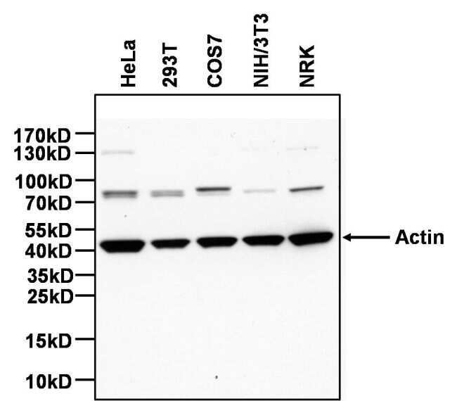
- Western blot analysis was performed on whole cell extracts (30 µg lysate) of A-431 (Lane 1), A549 (Lane 2), COS-7 (Lane 3), MDCK (Lane 4), NIH/3T3 (Lane 5), HT-29 (Lane 6), PC-12 (Lane 7), Neuro-2a (Lane 8), tissue extracts of Mouse Brain (Lane 9) and Rat Skeletal Muscle (Lane 10). The blot was probed with Actin Monoclonal Antibody (ACTN05 (C4)), Biotin (Product # MA5-11866, 1:5000 dilution) and detected by chemiluminescence using Poly-HRP Streptavidin (Product # N200, 1:10000 dilution). A 42 kDa band corresponding to Actin was observed across the cell lines and tissues tested.
- Conjugate
- Biotin

Supportive validation
- Submitted by
- Invitrogen Antibodies (provider)
- Main image
- Experimental details
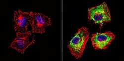
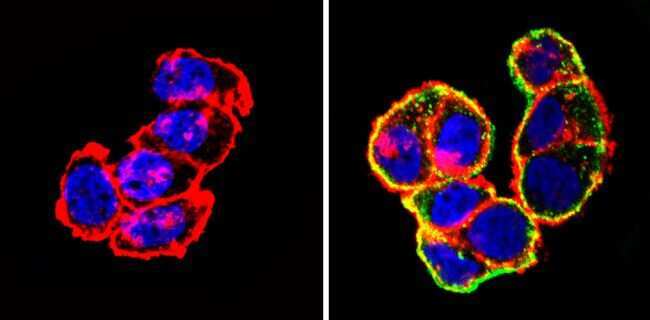
- Immunofluorescent analysis of Actin pan (green) showing staining in the cytoplasm of Hela cells (right) compared to a negative control without primary antibody (left). Formalin-fixed cells were permeabilized with 0.1% Triton X-100 in TBS for 5-10 minutes and blocked with 3% BSA-PBS for 30 minutes at room temperature. Cells were probed with an Actin pan monoclonal antibody (Product # MA5-11869) in 3% BSA-PBS at a dilution of 1:100 and incubated overnight at 4 ºC in a humidified chamber. Cells were washed with PBST and incubated with a DyLight-conjugated secondary antibody in PBS at room temperature in the dark. F-actin (red) was stained with a fluorescent red phalloidin and nuclei (blue) were stained with Hoechst or DAPI. Images were taken at a magnification of 60x.
- Conjugate
- Biotin
- Submitted by
- Invitrogen Antibodies (provider)
- Main image

- Experimental details
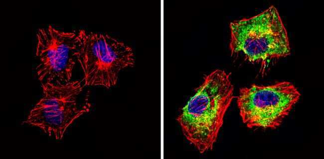
- Immunofluorescent analysis of Actin pan (green) showing staining in the cytoplasm of NIH-3T3 cells (right) compared to a negative control without primary antibody (left). Formalin-fixed cells were permeabilized with 0.1% Triton X-100 in TBS for 5-10 minutes and blocked with 3% BSA-PBS for 30 minutes at room temperature. Cells were probed with an Actin pan monoclonal antibody (Product # MA5-11869) in 3% BSA-PBS at a dilution of 1:100 and incubated overnight at 4 ºC in a humidified chamber. Cells were washed with PBST and incubated with a DyLight-conjugated secondary antibody in PBS at room temperature in the dark. F-actin (red) was stained with a fluorescent red phalloidin and nuclei (blue) were stained with Hoechst or DAPI. Images were taken at a magnification of 60x.
- Conjugate
- Biotin
- Submitted by
- Invitrogen Antibodies (provider)
- Main image

- Experimental details
- Immunofluorescent analysis of Actin pan (green) showing staining in the cytoplasm of T-47D cells (right) compared to a negative control without primary antibody (left). Formalin-fixed cells were permeabilized with 0.1% Triton X-100 in TBS for 5-10 minutes and blocked with 3% BSA-PBS for 30 minutes at room temperature. Cells were probed with an Actin pan monoclonal antibody (Product # MA5-11869) in 3% BSA-PBS at a dilution of 1:100 and incubated overnight at 4 ºC in a humidified chamber. Cells were washed with PBST and incubated with a DyLight-conjugated secondary antibody in PBS at room temperature in the dark. F-actin (red) was stained with a fluorescent red phalloidin and nuclei (blue) were stained with Hoechst or DAPI. Images were taken at a magnification of 60x.
- Conjugate
- Biotin
- Submitted by
- Invitrogen Antibodies (provider)
- Main image

- Experimental details
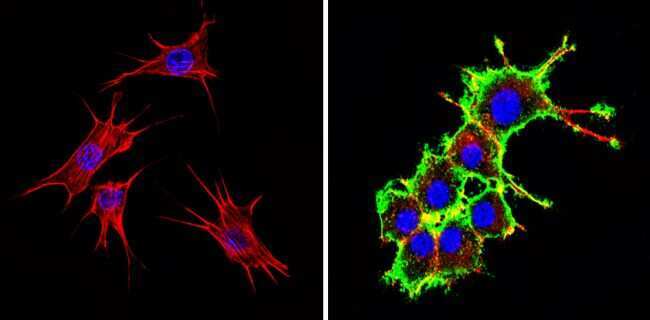
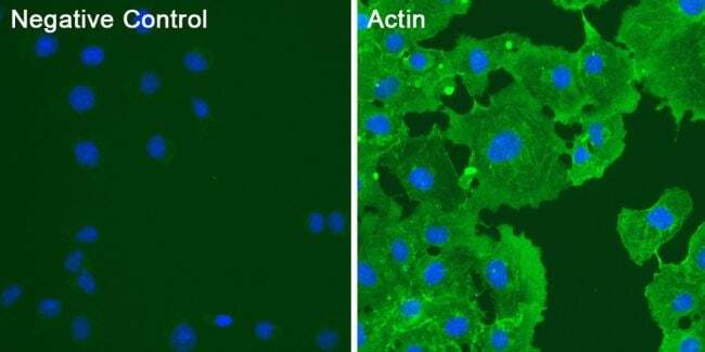
- Immunofluorescent analysis of actin (green) in COS7 cells. Cells were fixed and permeabilized with ice-cold methanol for 10 minutes at room temperature, and blocked with 0.3% BSA in PBS for 1 hour at room temperature. Cells were probed with a biotinylated pan actin monoclonal antibody (Product # MA5-11866) at a dilution of 1:50 (right panel) or incubated in blocking buffer as a negative control (left panel) overnight at 4°C, washed with PBS, and incubated with DyLight 488-conjugated Streptavidin (Product # 21832) at a dilution of 1:250 for 1 hour at room temperature. Nuclei (blue) were stained with DAPI (Product # 46190). Images were taken on a Thermo Scientific ToxInsight Instrument at 20X magnification.
- Conjugate
- Biotin

- Submitted by
- Invitrogen Antibodies (provider)
- Main image

- Experimental details
- Immunofluorescent analysis of actin (green) in COS7 cells. Cells were fixed and permeabilized with ice-cold methanol for 10 minutes at room temperature, and blocked with 0.3% BSA in PBS for 1 hour at room temperature. Cells were probed with a biotinylated pan actin monoclonal antibody (Product # MA5-11866) at a dilution of 1:50 (right panel) or incubated in blocking buffer as a negative control (left panel) overnight at 4°C, washed with PBS, and incubated with DyLight 488-conjugated Streptavidin (Product # 21832) at a dilution of 1:250 for 1 hour at room temperature. Nuclei (blue) were stained with DAPI (Product # 46190). Images were taken on a Thermo Scientific ToxInsight Instrument at 20X magnification.
- Conjugate
- Biotin
Supportive validation
- Submitted by
- Invitrogen Antibodies (provider)
- Main image
- Experimental details
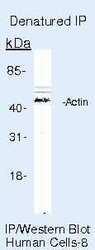
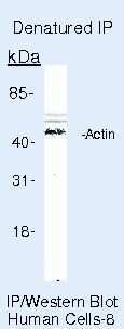
- Immunoprecipitation of Actin pan using Actin pan Monoclonal Antibody (Product # MA5-11866) on denatured Human HeLa Cells.
- Conjugate
- Biotin
- Submitted by
- Invitrogen Antibodies (provider)
- Main image

- Experimental details
- FIGURE 3: Protein degradation defects in the hac1Deltalpl1Delta mutant. (A) Levels of ubiquitin conjugates in whole-cell extracts of wild-type, lpl1Delta , hac1Delta , and hac1Deltalpl1Delta strains, as determined by SDS-PAGE followed by immunoblot with anti-ubiquitin antibody (top) or anti-Pgk1 antibody (bottom; loading control). Experiment was performed at 37degC. (B) Cycloheximide chase analysis of CPY* turnover in wild-type, lpl1Delta , hac1Delta , and hac1Deltalpl1Delta strains, as determined by SDS-PAGE followed by immunoblot with anti-HA antibody (top) or anti-actin antibody (bottom; loading control). Experiment was performed at 37degC. See Supplemental Figure S1 for quantitation. (C) Cycloheximide chase analysis of turnover of the cytoplasmic proteasome substrate Delta2-GFP, as determined by SDS-PAGE followed by immunoblot with anti-HA antibody (top) or anti-Pgk1 antibody (bottom; loading control). Experiment was performed at 37degC. (D) Growth of wild-type, lpl1Delta , hac1Delta , and hac1Deltalpl1Delta strains expressing galactose-inducible wild-type STE6 or the misfolded mutant ste6-Q1249X. Cells were spotted in threefold serial dilutions and cultured for 4 d at 35degC. (E) Levels of phosphorylated eIF2alpha (Ser-51) in whole-cell extracts of wild-type, lpl1Delta , hac1Delta , and hac1Deltalpl1Delta strains treated with sodium arsenite (0.2 mM), as determined by SDS-PAGE followed by immunoblot with anti-phospho-eIF2alpha antibody (top) or anti-Pgk1 antibody (bottom
- Conjugate
- Biotin

- Submitted by
- Invitrogen Antibodies (provider)
- Main image

- Experimental details
- Figure 3 Differences of RNA-binding motif protein 3 (RBM3) protein expression in various human breast cancer cell lines by western blot analysis. RBM3 protein is markedly increased in MCF-7 and T47D (luminal A subtype) and ZR-75-1 (luminal B subtype). On the other hand, RBM3 protein is significantly reduced in HCC1954 (HER2-enriched type) and BT-20 (basal-like subtype).
- Conjugate
- Biotin
