Explore
 Validate
 Learn
Learn Western blot
Antibody data
- Antibody Data
- Antigen structure
- References [0]
- Comments [0]
- Validations
- Western blot [4]
- Immunocytochemistry [3]
- Flow cytometry [2]
Submit
Validation data
Reference
Comment
Report error
- Product number
- 45-6200 - Provider product page

- Provider
- Invitrogen Antibodies
- Product name
- AIF Monoclonal Antibody (7F7AB10)
- Antibody type
- Monoclonal
- Antigen
- Other
- Description
- When performing IHC use heat mediated antigen retrieval before commencing with IHC staining protocol.
- Antibody clone number
- 7F7AB10
- Concentration
- 1 mg/mL
No comments: Submit comment
Supportive validation
- Submitted by
- Invitrogen Antibodies (provider)
- Main image

- Experimental details
- Western blot analysis of AIF was performed by loading 20 µg of K562 (lane1) and HeLa (lane2) cell lysates using Novex® NuPAGE® 4-12 % Bis-Tris gel (Product # NP0321BOX), XCell SureLock™ Electrophoresis System (Product # EI0002), Novex® Sharp Pre-Stained Protein Standard (LC5800), and iBlot® Dry Blotting System (IB21001). Proteins were transferred to a nitrocellulose membrane and blocked with 5 % skim milk for 1 hour at room temperature. AIF was detected at ~63 kDa using AIF Mouse Monoclonal Antibody (Product # 45-6200) at 1 µg - 3 µg/mL in 2.5 % skim milk at 4°C overnight on a rocking platform. Goat Anti-Mouse IgG - HRP Secondary Antibody (Product # 62-6520) at 1:4000 dilution was used and chemiluminescent detection was performed using Pierce™ ECL Western Blotting Substrate (Product # 32106).
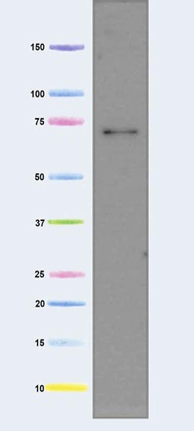
- Submitted by
- Invitrogen Antibodies (provider)
- Main image
- Experimental details
- Western blot analysis of AIF in isolated mitochondria using a PDH Monoclonal antibody (Product # 45-6600) at a concentration of 5 µg/mL. Isolated mitochondria from Human heart at 5 µg. Predicted band size is 67 kDa.
- Submitted by
- Invitrogen Antibodies (provider)
- Main image

- Experimental details
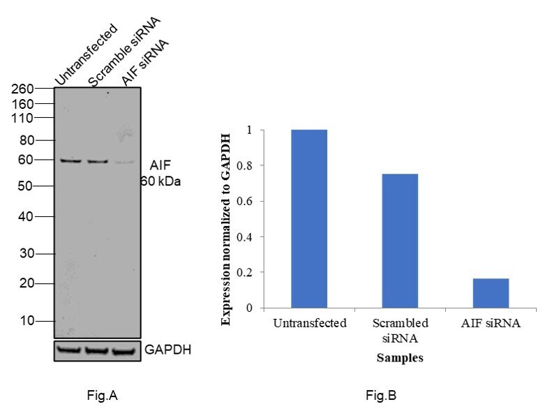
- Knockdown of AIF was achieved by transfecting HeLa with AIF specific siRNAs (Silencer® select Product # S17442, S17440). Western blot analysis (Fig. a) was performed using Whole cell extracts from the AIF untransfected cells (lane 1), non-targeting scrambled siRNA transfected cells (lane 2) and knockdown cells (lane 3). The blot was probed with AIF Monoclonal Antibody (7F7AB10) (Product # 45-6200, 1:500 dilution ) and Goat anti-Mouse IgG (H+L) Superclonal™ Recombinant Secondary Antibody, HRP (Product # A28177, 1:4000 dilution). Densitometric analysis of this western blot is shown in histogram (Fig. b). Decrease in signal upon siRNA mediated knock down confirms that antibody is specific to AIF.
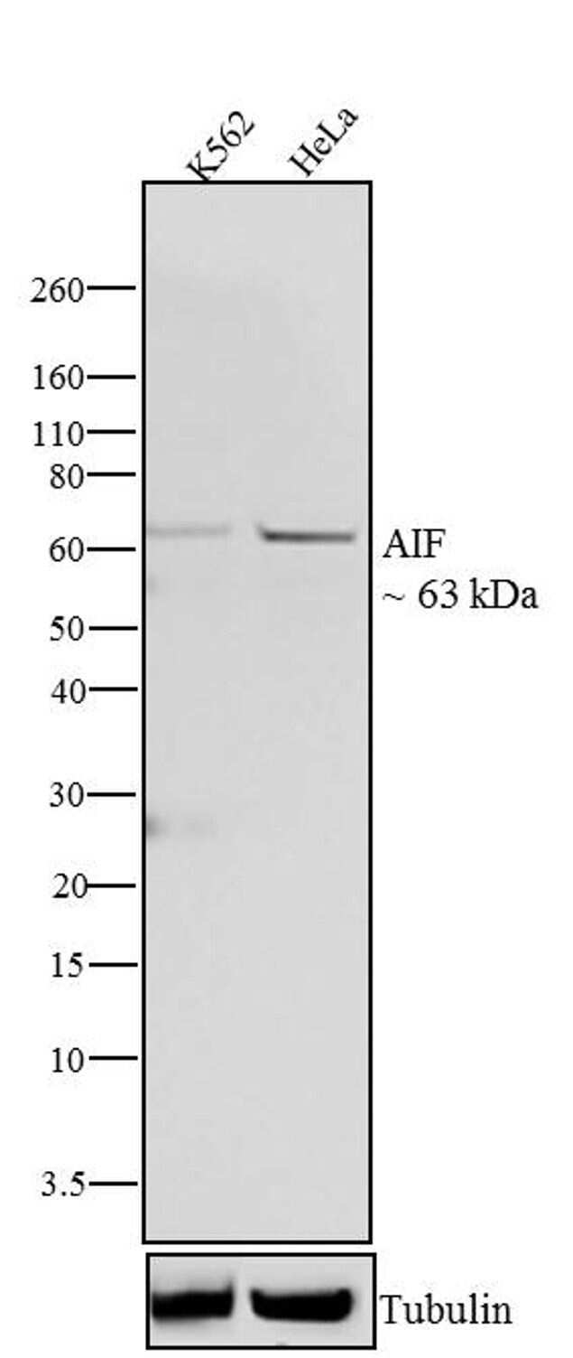
- Submitted by
- Invitrogen Antibodies (provider)
- Main image
- Experimental details
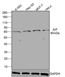
- Western blot was performed using Anti-AIF Monoclonal Antibody (7F7AB10)(Product # 45-6200) and a 60kDa band corresponding to AIF was observed across cell lines tested. Whole cell extracts (30 µg lysate) of K-562 (Lane 1), Hep G2 (Lane 2), MCF7 (Lane 3), HeLa (Lane 4) were electrophoresed using NuPAGE™ 4-12% Bis-Tris Protein Gel (Product # NP0322BOX). Resolved proteins were then transferred onto a Nitrocellulose membrane (Product # IB23001) by iBlot® 2 Dry Blotting System (Product # IB21001). The blot was probed with the primary antibody (1:500 Dilution) and detected by chemiluminescence with Goat anti-Mouse IgG (H+L) Superclonal™ Recombinant Secondary Antibody, HRP (Product # A28177, 1:4000 dilution) using the iBright FL 1000 (Product # A32752). Chemiluminescent detection was performed using Novex® ECL Chemiluminescent Substrate Reagent Kit (Product # WP20005).

Supportive validation
- Submitted by
- Invitrogen Antibodies (provider)
- Main image

- Experimental details
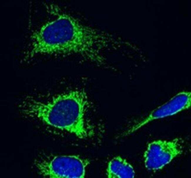
- Immunofluorescent analysis of AIF in HeLa cells using an AIF Monoclonal antibody (Product # 45-6600) at 5 µg/mL. HeLa cells were fixed in 4% paraformaldehyde and permeabilized with Triton X-100. The secondary antibody was Alexa Fluor® 488 goat anti-mouse IgG (H+L) used at a 1/1000 dilution for 1 hour, as seen in green. DAPI was used to stain the cell nuclei blue. AIF is localized mainly in the mitochondria.
- Submitted by
- Invitrogen Antibodies (provider)
- Main image
- Experimental details
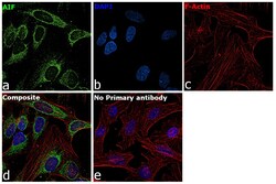
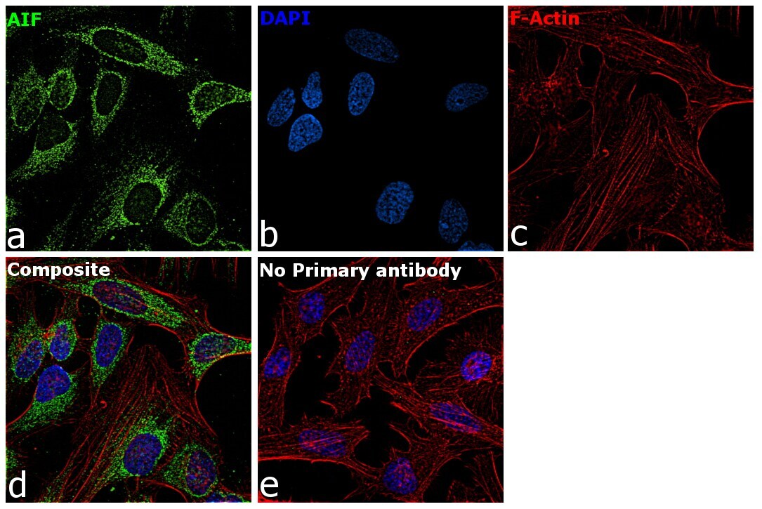
- Immunofluorescence analysis of AIF was performed using 70% confluent log phase HeLa cells. The cells were fixed with 4% paraformaldehyde for 10 minutes, permeabilized with 0.1% Triton™ X-100 for 15 minutes, and blocked with 2% BSA for 45 minutes at room temperature. The cells were labeled with AIF Monoclonal Antibody (7F7AB10) (Product # 45-6200) at 5 µg in 0.1% BSA, incubated at 4 degree celsius overnight and then labeled with Goat anti-Mouse IgG (H+L) Superclonal™ Recombinant Secondary Antibody, Alexa Fluor® 488 conjugate (Product # A28175), (1:2000 dilution), for 45 minutes at room temperature (Panel a: Green). Nuclei (Panel b:Blue) were stained with ProLong™ Diamond Antifade Mountant with DAPI (Product # P36962). F-actin (Panel c: Red) was stained with Rhodamine Phalloidin (Product # R415, 1:300 dilution). Panel d represents the merged image showing mitochondria localization. Panel e represents control cells with no primary antibody to assess background. The images were captured at 60x magnification.
- Submitted by
- Invitrogen Antibodies (provider)
- Main image

- Experimental details
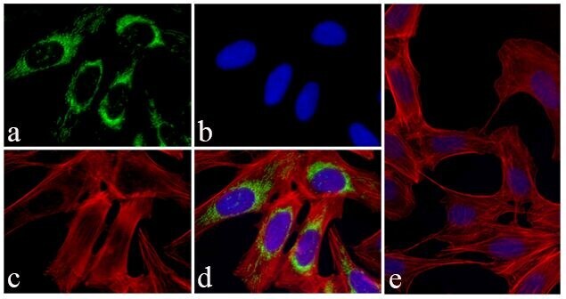
- Immunofluorescent analysis of AIF was done on 70% confluent log phase HeLa cells. The cells were fixed with 4% paraformaldehyde for 15 minutes; permeabilized with 0.25% Triton™ X-100 for 10 minutes followed by blocking with 5% BSA for 1 hour at room temperature. The cells were incubated with AIF Mouse Monoclonal Antibody (Product # 45-6200) at 1 µg - 2 µg in 1% BSA and incubated for 3 hours at room temperature and then labeled with Alexa Fluor® 488 Rabbit Anti-Mouse IgG Secondary Antibody (Product # A-11059) at a dilution of 1:400 for 30 minutes at room temperature (Panel a: green). Nuclei (Panel b: blue) were stained with SlowFade® Gold Antifade Mountant with DAPI (Product # S36938). F-actin (Panel c: red) was stained with Alexa Fluor® 594 Phalloidin (Product # A12381). Panel d is a merged image showing cytoplasmic localization of AIF. Panel e shows no primary antibody control. The images were captured at 20X magnification.
Supportive validation
- Submitted by
- Invitrogen Antibodies (provider)
- Main image
- Experimental details
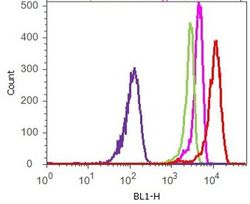
- Flow cytometry analysis of AIF was done on Jurkat cells. Cells were fixed with 70% ethanol for 10 minutes, permeabilized with 0.25% Triton™ X-100 for 20 minutes, and blocked with 5% BSA for 1 hour at room temperature. Cells were labeled with AIF Mouse Monoclonal Antibody (456200, red histogram) or with mouse isotype control (pink histogram) at 2 µg - 4 µg/million cells in 2.5% BSA. After incubation at room temperature for 2 - 3 hours, the cells were labeled with Alexa Fluor® 488 Rabbit Anti-Mouse Secondary Antibody (A11059) at a dilution of 1:400 for 30 minutes at room temperature. The representative 10,000 cells were acquired and analyzed for each sample using an Attune® Acoustic Focusing Cytometer. The purple histogram represents unstained control cells and the green histogram represents no-primary-antibody control.

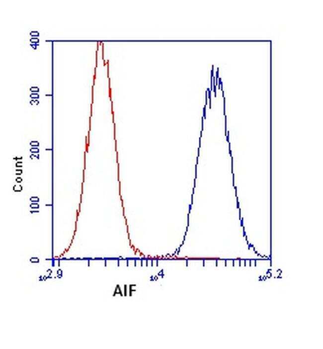
- Submitted by
- Invitrogen Antibodies (provider)
- Main image

- Experimental details
- Flow cytometric analysis of AIF in HL-60 cells using an AIF Monoclonal antibody (Product # 45-6200) at 1 µg/mL, as shown in blue. Isotype control antibody is shown in red.