 Explore
 Validate
 Learn
LearnPA5-19947
antibody from Invitrogen Antibodies
Targeting: AIFM1
AIF, AUNX1, CMTX4, DFNX5, NAMSD, PDCD8
 Western blot
Antibody data
- Antibody Data
- Antigen structure
- References [0]
- Comments [0]
- Validations
- Western blot [4]
- Immunocytochemistry [3]
- Immunohistochemistry [1]
Submit
Validation data
Reference
Comment
Report error
- Product number
- PA5-19947 - Provider product page

- Provider
- Invitrogen Antibodies
- Product name
- AIF Polyclonal Antibody
- Antibody type
- Polyclonal
- Antigen
- Synthetic peptide
- Description
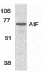
- In Western blot applications, this antibody detects a band at ~67kDa. A suggested positive control is K562 cell lysate.
- Concentration
- 1 mg/mL
No comments: Submit comment
Supportive validation
- Submitted by
- Invitrogen Antibodies (provider)
- Main image
- Experimental details
- Western blot analysis of K562 cell lysate using a AIF polyclonal antibody (Product # PA5-19947) at 1 µg/mL.

- Submitted by
- Invitrogen Antibodies (provider)
- Main image

- Experimental details
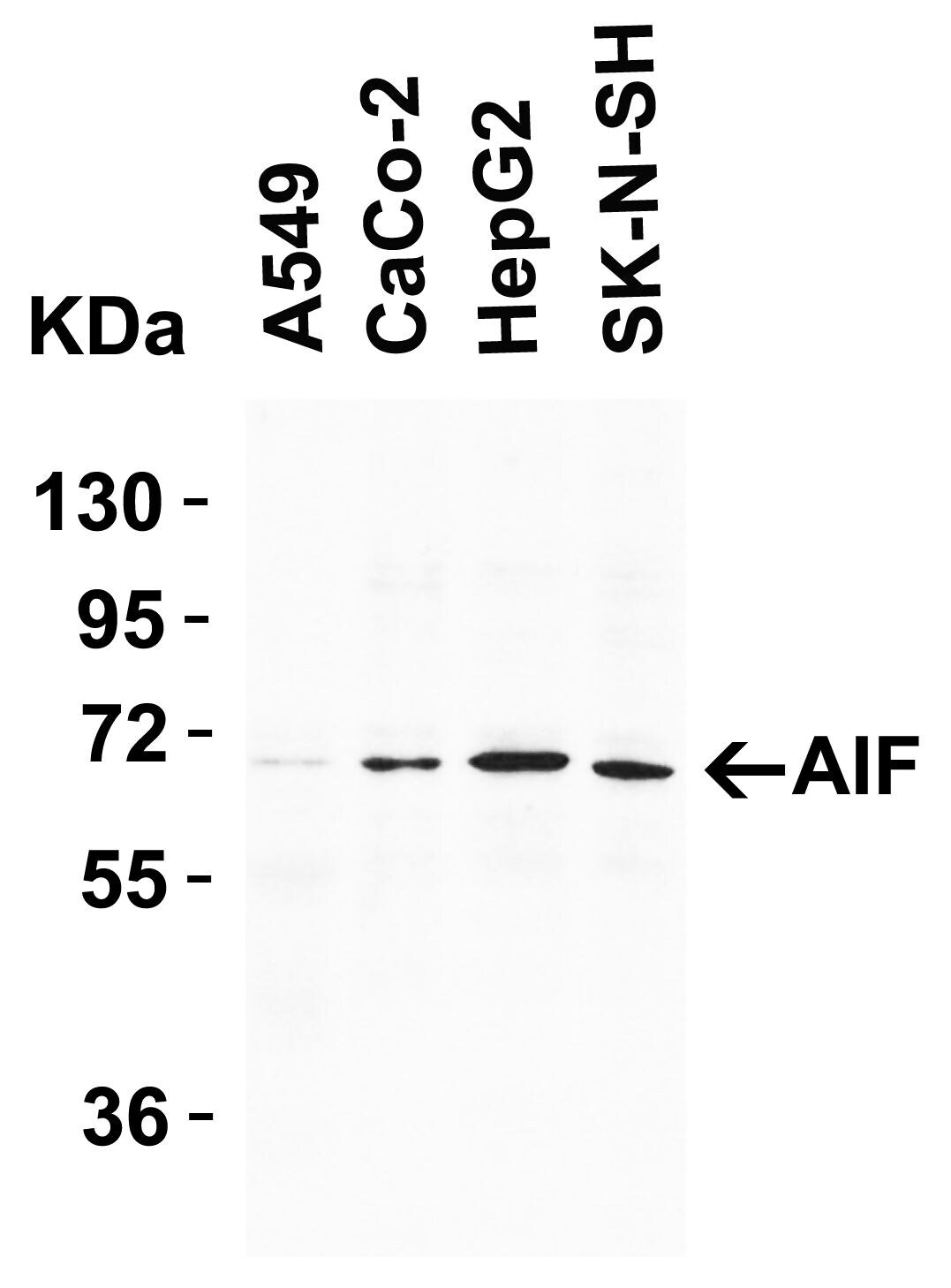
- Western Blot Validation in Human Cell Lines. Loading: 15 µg of lysates per lane. Antibodies: AIF Polyclonal Antibody (Product # PA5-19947) (1 µg/mL), 1h incubation at RT in 0.05 NFDM/TBST. Secondary: Goat anti-rabbit IgG HRP conjugate at 1:10,000 dilution.
- Submitted by
- Invitrogen Antibodies (provider)
- Main image

- Experimental details
- Knockdown of AIF was achieved by transfecting HeLa with AIF specific siRNAs (Silencer® select Product # S17442, S17440). Western blot analysis (Fig. a) was performed using Whole cell extracts from the AIF untransfected cells (lane 1), non-targeting scrambled siRNA transfected cells (lane 2) and knockdown cells (lane 3). The blot was probed with AIF Polyclonal Antibody (Product # PA5-19947, 1:1000 dilution ) and Goat anti-Rabbit IgG (H+L) Superclonal™ Recombinant Secondary Antibody, HRP (Product # A27036, 1:4000 dilution). Densitometric analysis of this western blot is shown in histogram (Fig. b). Decrease in signal upon siRNA mediated knock down confirms that antibody is specific to AIF.

- Submitted by
- Invitrogen Antibodies (provider)
- Main image

- Experimental details
- Western blot was performed using Anti-AIF Polyclonal Antibody(Product # PA5-19947) and a 60kDa band corresponding to AIF was observed across cell lines tested. Whole cell extracts (30 µg lysate) of K-562 (Lane 1), Hep G2 (Lane 2), MCF7 (Lane 3), HeLa (Lane 4) were electrophoresed using NuPAGE™ 4-12% Bis-Tris Protein Gel (Product # NP0322BOX). Resolved proteins were then transferred onto a Nitrocellulose membrane (Product # IB23001) by iBlot® 2 Dry Blotting System (Product # IB21001). The blot was probed with the primary antibody (1:1000 Dilution) and detected by chemiluminescence with Goat anti-Rabbit IgG (H+L) Superclonal™ Recombinant Secondary Antibody, HRP (Product # A27036,1:4000 dilution) using the iBright FL 1000 (Product # A32752). Chemiluminescent detection was performed using Novex® ECL Chemiluminescent Substrate Reagent Kit (Product # WP20005).

Supportive validation
- Submitted by
- Invitrogen Antibodies (provider)
- Main image
- Experimental details
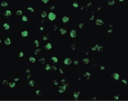
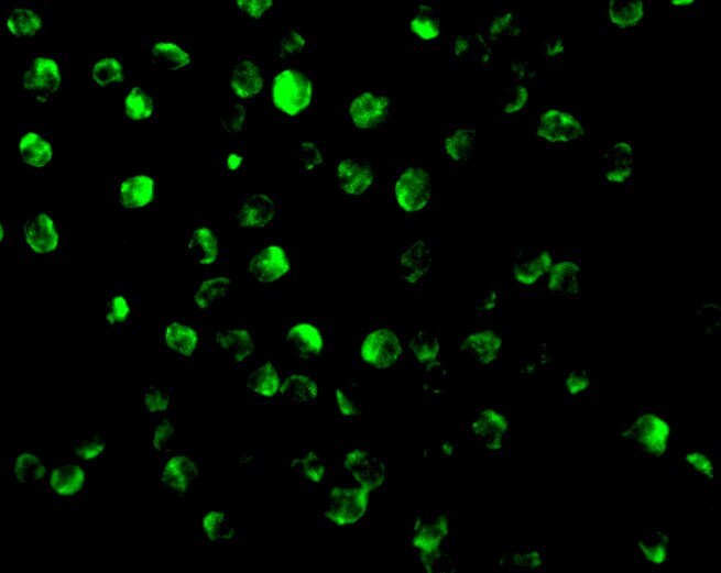
- Immunofluorescent analysis of K562 cells using a AIF polyclonal antibody (Product # PA5-19947) at a 20 µg/mL dilution.
- Submitted by
- Invitrogen Antibodies (provider)
- Main image

- Experimental details
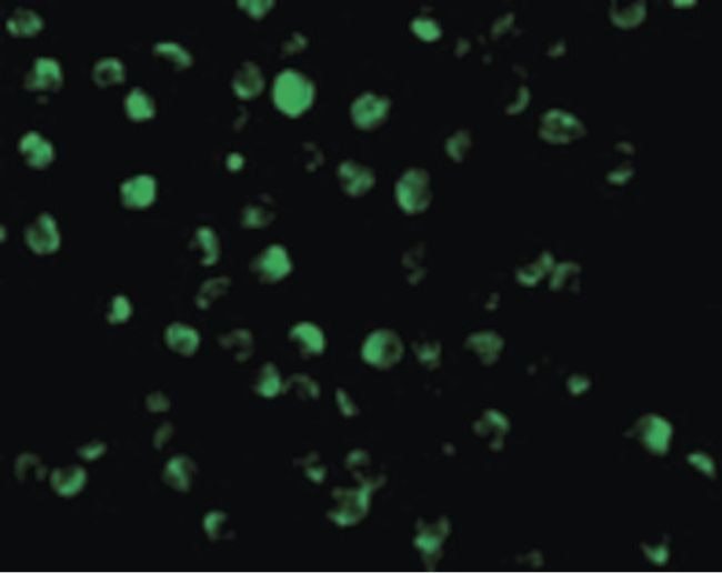
- Immunofluorescent analysis of 4% paraformaldehyde-fixed K562 Cells labeling AIF with AIF Polyclonal Antibody (Product # PA5-19947) at 20 µg/mL, followed by goat anti-rabbit IgG secondary antibody at 1:500 dilution (green).
- Submitted by
- Invitrogen Antibodies (provider)
- Main image
- Experimental details
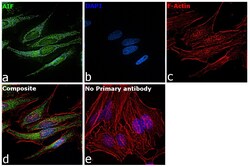
- Immunofluorescence analysis of AIF was performed using 70% confluent log phase HeLa cells. The cells were fixed with 4% paraformaldehyde for 10 minutes, permeabilized with 0.1% Triton™ X-100 for 15 minutes, and blocked with 2% BSA for 45 minutes at room temperature. The cells were labeled with AIF Polyclonal Antibody (Product # PA5-19947) at 5 µg in 0.1% BSA, incubated at 4 degree celsius overnight and then labeled with Goat anti-Rabbit IgG (H+L) Superclonal™ Recombinant Secondary Antibody, Alexa Fluor® 488 conjugate (Product # A27034), (1:2000 dilution), for 45 minutes at room temperature (Panel a: Green). Nuclei (Panel b:Blue) were stained with SlowFade® Gold Antifade Mountant with DAPI (Product # S36938). F-actin (Panel c: Red) was stained with Rhodamine Phalloidin (Product # R415, 1:300 dilution). Panel d represents the merged image showing mitochondrial localization. Panel e represents control cells with no primary antibody to assess background. The images were captured at 60x magnification.

Supportive validation
- Submitted by
- Invitrogen Antibodies (provider)
- Main image

- Experimental details
- Immunocytochemistry staining of Jurkat cells using a AIF polyclonal antibody (Product # PA5-19947) at a 2 µg/mL dilution.
