 Explore
 Validate
 Learn
LearnPA5-19953
antibody from Invitrogen Antibodies
Targeting: AIFM1
AIF, AUNX1, CMTX4, DFNX5, NAMSD, PDCD8
 Western blot
Antibody data
- Antibody Data
- Antigen structure
- References [0]
- Comments [0]
- Validations
- Western blot [6]
- Immunocytochemistry [1]
- Immunohistochemistry [1]
Submit
Validation data
Reference
Comment
Report error
- Product number
- PA5-19953 - Provider product page

- Provider
- Invitrogen Antibodies
- Product name
- AIF Polyclonal Antibody
- Antibody type
- Polyclonal
- Antigen
- Synthetic peptide
- Description
- In Western blot applications, this antibody detects a band at ~67kDa. A suggested positive control is K562 cell lysate. PA5-19953 can be used with blocking peptide PEP-0078.
- Reactivity
- Human, Mouse, Rat
- Host
- Rabbit
- Isotype
- IgG
- Vial size
- 100 µg
- Concentration
- 1 mg/mL
- Storage
- Maintain refrigerated at 2-8°C for up to 3 months. For long term storage store at -20°C
No comments: Submit comment
Supportive validation
- Submitted by
- Invitrogen Antibodies (provider)
- Main image
- Experimental details
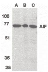
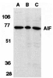
- Western blot analysis of K562 cell lysate (A), rat heart (B), and mouse heart (C) tissue lysates using a AIF polyclonal antibody (Product # PA5-19953) at 1 µg/mL.
- Submitted by
- Invitrogen Antibodies (provider)
- Main image
- Experimental details
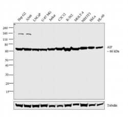
- Western blot analysis was performed on whole cell extracts (30 µg lysate) of HepG2 (Lane 1), A549 (Lane 2), LNCaP (Lane 3), U-87 MG (Lane 4), Jurkat (Lane 5), C2C12 (Lane 6), K-562 (Lane 7), MOLT-4 (Lane 8), NIH/3T3 (Lane 9), HeLa (Lane 10) and HL-60 (Lane 11). The blot was probed with Anti-AIF Polyclonal Antibody (Product # PA5-19953, 1:500 dilution) and detected by chemiluminescence using Goat anti-Rabbit IgG (H+L) Superclonal™ Secondary Antibody, HRP conjugate (Product # A27036, 0.25 µg/mL, 1:4000 dilution). A 66 kDa band corresponding to AIF was observed across the cell lines tested.

- Submitted by
- Invitrogen Antibodies (provider)
- Main image
- Experimental details
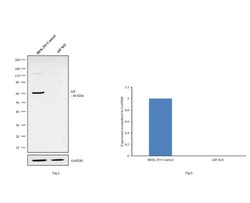
- Western blot analysis of AIF (Fig. a) was performed by loading 20 µg of HEK-293 Control (lane 1), HEK-293 AIF knockout (lane 2) whole cell extracts. AIF was detected at 66 kDa using AIF Monoclonal Antibody (4E7) (Product # PA5-19953, 1 µg/mL) and Goat anti-Rabbit IgG (H+L) Superclonal™ Secondary Antibody, HRP conjugate (Product # A27036, 0.25µg/mL, 1:4000 dilution). Densitometric analysis of this western blot is shown in histogram (Fig. b). Loss of signal in CRISPR mediated knockout (KO) confirms that antibody is specific to AIF.

- Submitted by
- Invitrogen Antibodies (provider)
- Main image
- Experimental details
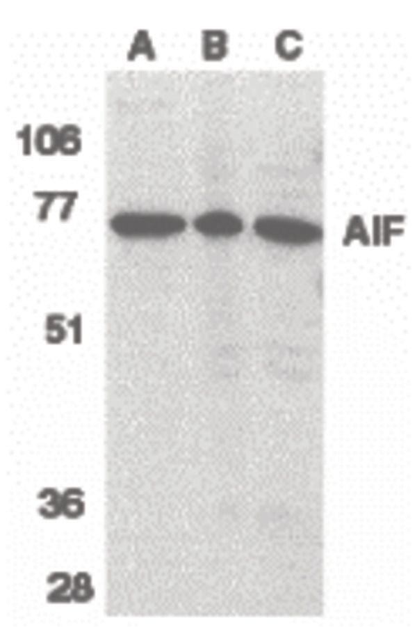
- Western Blot Validation in Different Species. Loading: 15 µg of lysates per lane. Antibodies: AIF Polyclonal Antibody (Product # PA5-19953) (1 µg/mL), 1h incubation at RT in 0.05 NFDM/TBST. Secondary: Goat anti-rabbit IgG HRP conjugate at 1:10,000 dilution. Lane A: Human K562 cells Lane B: Rat heart Lane C: Mouse heart

- Submitted by
- Invitrogen Antibodies (provider)
- Main image
- Experimental details
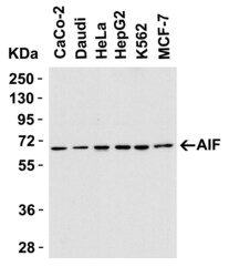
- Western Blot Validation in Human Cell Lines. Loading: 15 µg of lysates per lane. Antibodies: AIF Polyclonal Antibody (Product # PA5-19953) (1 µg/mL), 1h incubation at RT in 0.05 NFDM/TBST. Secondary: Goat anti-rabbit IgG HRP conjugate at 1:10,000 dilution.

- Submitted by
- Invitrogen Antibodies (provider)
- Main image
- Experimental details
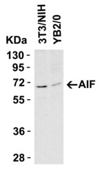
- Western Blot Validation in Mouse and Rat Cell Lines. Loading: 15 µg of lysates per lane. Antibodies: AIF Polyclonal Antibody (Product # PA5-19953) (1 µg/mL), 1h incubation at RT in 0.05 NFDM/TBST. Secondary: Goat anti-rabbit IgG HRP conjugate at 1:10,000 dilution.

Supportive validation
- Submitted by
- Invitrogen Antibodies (provider)
- Main image

- Experimental details
- Immunofluorescence analysis of AIF was performed using 70% confluent log phase Hep G2 cells. The cells were fixed with 4% paraformaldehyde for 10 minutes, permeabilized with 0.1% Triton™ X-100 for 10 minutes, and blocked with 1% BSA for 1 hour at room temperature. The cells were labeled with AIF Polyclonal Antibody (Product # PA5-19953) at 1:250 dilution in 0.1% BSA and incubated overnight at 4 degree and then labeled with Goat anti-Rabbit IgG (H+L) Superclonal™ Secondary Antibody, Alexa Fluor® 488 conjugate (Product # A27034) at a dilution of 1:2000 for 45 minutes at room temperature (Panel a: green). Nuclei (Panel b: blue) were stained with ProLong™ Diamond Antifade Mountant with DAPI (Product # P36962). Panel c represents mitochondrial staining using MitoTracker® Red CMXRos (Product # M7512). Panel d is a merged image of Panels a, b and c clearly demonstrating co-localization of AIF with mitotracker which specifically binds to the mitochondria. Panel e represents control cells with no primary antibody to assess background. The images were captured at 60X magnification.

Supportive validation
- Submitted by
- Invitrogen Antibodies (provider)
- Main image
- Experimental details
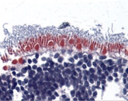
- Immunohistochemical analysis of paraffin-embedded human retina tissue using AIF Polyclonal Antibody (Product # PA5-19953) at 10 µg/mL. Tissue was fixed with formaldehyde and blocked with 0.1 serum for 1 h at RT; antigen retrieval was by heat mediation with a citrate buffer (pH6). Samples were incubated with primary antibody overnight at 4˚ C. A goat anti-rabbit IgG H&L (HRP) at 1/250 was used as secondary. Counter stained with Hematoxylin.
