Explore
 Validate
 Learn
Learn Western blot
Antibody data
- Antibody Data
- Antigen structure
- References [6]
- Comments [0]
- Validations
- Western blot [3]
- Immunocytochemistry [3]
Submit
Validation data
Reference
Comment
Report error
- Product number
- AF1457 - Provider product page

- Provider
- R&D Systems
- Product name
- Human/Mouse/Rat AIF Antibody
- Antibody type
- Polyclonal
- Description
- Antigen Affinity-purified. Detects human, mouse, and rat mitochondria-processed AIF.
- Reactivity
- Human, Mouse, Rat
- Host
- Rabbit
- Conjugate
- Unconjugated
- Antigen sequence
O95831- Isotype
- IgG
- Vial size
- 100 ug
- Concentration
- LYOPH
- Storage
- Use a manual defrost freezer and avoid repeated freeze-thaw cycles. 12 months from date of receipt, -20 to -70 °C as supplied. 1 month, 2 to 8 °C under sterile conditions after reconstitution. 6 months, -20 to -70 °C under sterile conditions after reconstitution.
Submitted references Glyceraldehyde-3-phosphate Dehydrogenase (GAPDH) Aggregation Causes Mitochondrial Dysfunction during Oxidative Stress-induced Cell Death.
Delivery of a survivin promoter-driven antisense survivin-expressing plasmid DNA as a cancer therapeutic: a proof-of-concept study.
Novel Biomarker Proteins in Chronic Lymphocytic Leukemia: Impact on Diagnosis, Prognosis and Treatment.
Diet-induced obesity exacerbates auditory degeneration via hypoxia, inflammation, and apoptosis signaling pathways in CD/1 mice.
The regulatory roles of apoptosis-inducing factor in the formation and regression processes of ocular neovascularization.
Critical involvement of extracellular ATP acting on P2RX7 purinergic receptors in photoreceptor cell death.
Nakajima H, Itakura M, Kubo T, Kaneshige A, Harada N, Izawa T, Azuma YT, Kuwamura M, Yamaji R, Takeuchi T
The Journal of biological chemistry 2017 Mar 17;292(11):4727-4742
The Journal of biological chemistry 2017 Mar 17;292(11):4727-4742
Delivery of a survivin promoter-driven antisense survivin-expressing plasmid DNA as a cancer therapeutic: a proof-of-concept study.
Lin KY, Cheng SM, Tsai SL, Tsai JY, Lin CH, Cheung CH
OncoTargets and therapy 2016;9:2601-13
OncoTargets and therapy 2016;9:2601-13
Novel Biomarker Proteins in Chronic Lymphocytic Leukemia: Impact on Diagnosis, Prognosis and Treatment.
Admoni-Elisha L, Nakdimon I, Shteinfer A, Prezma T, Arif T, Arbel N, Melkov A, Zelichov O, Levi I, Shoshan-Barmatz V
PloS one 2016;11(4):e0148500
PloS one 2016;11(4):e0148500
Diet-induced obesity exacerbates auditory degeneration via hypoxia, inflammation, and apoptosis signaling pathways in CD/1 mice.
Hwang JH, Hsu CJ, Yu WH, Liu TC, Yang WS
PloS one 2013;8(4):e60730
PloS one 2013;8(4):e60730
The regulatory roles of apoptosis-inducing factor in the formation and regression processes of ocular neovascularization.
Hisatomi T, Nakao S, Murakami Y, Noda K, Nakazawa T, Notomi S, Connolly E, She H, Almulki L, Ito Y, Vavvas DG, Ishibashi T, Miller JW
The American journal of pathology 2012 Jul;181(1):53-61
The American journal of pathology 2012 Jul;181(1):53-61
Critical involvement of extracellular ATP acting on P2RX7 purinergic receptors in photoreceptor cell death.
Notomi S, Hisatomi T, Kanemaru T, Takeda A, Ikeda Y, Enaida H, Kroemer G, Ishibashi T
The American journal of pathology 2011 Dec;179(6):2798-809
The American journal of pathology 2011 Dec;179(6):2798-809
No comments: Submit comment
Supportive validation
- Submitted by
- R&D Systems (provider)
- Main image
- Experimental details
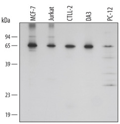
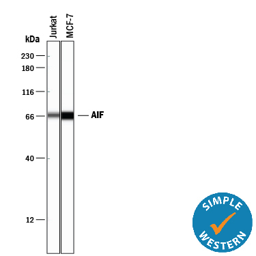
- Detection of Human/Mouse/Rat AIF by Western Blot. Western blot shows lysates of MCF-7 human breast cancer cell line, Jurkat human acute T cell leukemia cell line, CTLL-2 mouse cytotoxic T cell line, DA3 mouse myeloma cell line, and PC-12 rat adrenal pheochromocytoma cell line. PVDF membrane was probed with 0.1 µg/mL of Rabbit Anti-Human/Mouse/Rat AIF Antigen Affinity-purified Polyclonal Antibody (Catalog # AF1457) followed by HRP-conjugated Anti-Rabbit IgG Secondary Antibody (Catalog # HAF008). A specific band was detected for AIF at approximately 67 kDa (as indicated). This experiment was conducted under reducing conditions and using Immunoblot Buffer Group 2.
- Submitted by
- R&D Systems (provider)
- Main image
- Experimental details
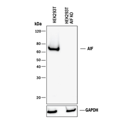
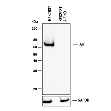
- Western Blot Shows Human AIF Specificity by Using Knockout Cell Line. Western blot shows lysates of HEK293T human embryonic kidney parental cell line and AIF knockout HEK293T cell line (KO). PVDF membrane was probed with 0.1 µg/mL of Rabbit Anti-Human/Mouse/Rat AIF Antigen Affinity-purified Polyclonal Antibody (Catalog # AF1457) followed by HRP-conjugated Anti-Rabbit IgG Secondary Antibody (Catalog # HAF008). A specific band was detected for AIF at approximately 65 kDa (as indicated) in the parental HEK293T cell line, but is not detectable in knockout HEK293T cell line. GAPDH (Catalog # AF5718) is shown as a loading control. This experiment was conducted under reducing conditions and using Immunoblot Buffer Group 1.
- Submitted by
- R&D Systems (provider)
- Main image
- Experimental details
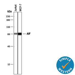
- Detection of Human AIF by Simple WesternTM. Simple Western lane view shows lysates of Jurkat human acute T cell leukemia cell line and MCF-7 human breast cancer cell line, loaded at 0.2 mg/mL. A specific band was detected for AIF at approximately 69 kDa (as indicated) using 1 µg/mL of Rabbit Anti-Human/Mouse/Rat AIF Antigen Affinity-purified Polyclonal Antibody (Catalog # AF1457). This experiment was conducted under reducing conditions and using the 12-230 kDa separation system.
Supportive validation
- Submitted by
- R&D Systems (provider)
- Main image
- Experimental details
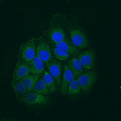
- AIF in MCF-7 Human Cell Line. Apoptosis Inducing Factor (AIF) was detected in immersion fixed MCF-7 human breast cancer cell line using Rabbit Anti-Human/Mouse/Rat AIF Antigen Affinity-purified Polyclonal Antibody (Catalog # AF1457) at 10 µg/mL for 3 hours at room temperature. Cells were stained using the NorthernLights™ 493-conjugated Anti-Rabbit IgG Secondary Antibody (green; Catalog # NL006) and counterstained with DAPI (blue). View our protocol for Fluorescent ICC Staining of Cells on Coverslips.

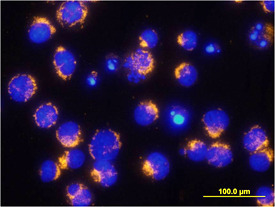
- Submitted by
- R&D Systems (provider)
- Main image

- Experimental details
- AIF in Jurkat Human Cell Line. AIF was detected in immersion fixed staurosporine-stimulated Jurkat human acute T cell leukemia cell line using Rabbit Anti-Human/Mouse/Rat AIF Antigen Affinity-purified Polyclonal Antibody (Catalog # AF1457) at 10 µg/mL for 3 hours at room temperature. Cells were stained using the Northern-Lights™ 557-conjugated Anti-Rabbit IgG Secondary Antibody (yellow; Catalog # NL004) and counter-stained with DAPI (blue). View our protocol for Fluorescent ICC Staining of Non-adherent Cells.
- Submitted by
- R&D Systems (provider)
- Main image

- Experimental details
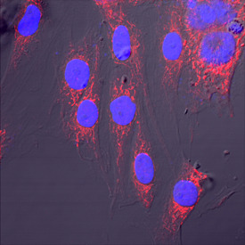
- AIF in HeLa Human Cell Line. AIF was detected in immersion fixed HeLa human cervical epithelial carcinoma cell line using Rabbit Anti-Human/Mouse/Rat AIF Antigen Affinity-purified Polyclonal Antibody (Catalog # AF1457) at 15 µg/mL for 3 hours at room temperature. Cells were stained using the NorthernLights™ 557-conjugated Anti-Sheep IgG Secondary Antibody (red; Catalog # NL010) and counterstained with DAPI (blue). Specific staining was localized to mitochondria. View our protocol for Fluorescent ICC Staining of Cells on Coverslips.