Explore
 Validate
 Learn
Learn Western blot
Antibody data
- Antibody Data
- Antigen structure
- References [0]
- Comments [0]
- Validations
- Western blot [3]
- Immunocytochemistry [3]
- Immunohistochemistry [7]
- Flow cytometry [1]
Submit
Validation data
Reference
Comment
Report error
- Product number
- R31583 - Provider product page

- Provider
- NSJ Bioreagents
- Product name
- Caveolin-1 Antibody
- Antibody type
- Polyclonal
- Description
- This highly specific Caveolin-1 antibody is suitable for use in Western blot/Immunohistochemistry/Immunohistochemistry/Immunofluorescence/Flow cytometry applications with human, mouse and rat samples.
- Reactivity
- Human, Mouse, Rat
- Host
- Rabbit
- Conjugate
- Unconjugated
- Vial size
- 100 ug
- Concentration
- 0.5mg/ml if reconstituted with 0.2ml sterile DI water
- Storage
- After reconstitution, the Caveolin-1 antibody can be stored for up to one month at 4oC. For long-term, aliquot and store at -20oC. Avoid repeated freezing and thawing.
No comments: Submit comment
Supportive validation
- Submitted by
- NSJ Bioreagents (provider)
- Main image
- Experimental details
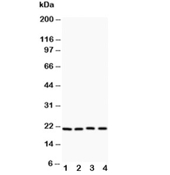
- Western blot testing of Caveolin-1 antibody and Lane 1: HeLa; 2: HT1080; 3: human placenta; 4: A431

- Submitted by
- NSJ Bioreagents (provider)
- Main image
- Experimental details
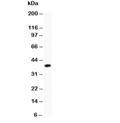
- Western blot testing of Caveolin-1 antibody and recombinant human protein (0.5ng)

- Submitted by
- NSJ Bioreagents (provider)
- Main image
- Experimental details
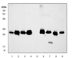
- Western blot testing of 1) human HeLa, 2) human A549, 3) human placenta, 4) human A431, 5) human HL60, 6) rat ovary, 7) rat heart, 8) mouse ovary and 9) mouse heart tissue lysate with Caveolin-1 antibody. Predicted molecular weight ~21 kDa.

Supportive validation
- Submitted by
- NSJ Bioreagents (provider)
- Main image

- Experimental details
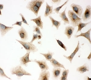
- ICC testing of A549 cells with Caveolin-1 antibody.
- Submitted by
- NSJ Bioreagents (provider)
- Main image
- Experimental details
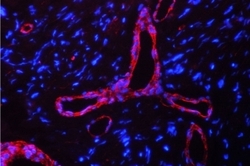
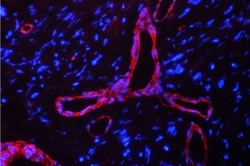
- Immunofluorescent staining of FFPE human glioma tissue with Calveolin-1 antibody (red) and DAPI nuclear counterstain (blue). HIER: boil tissue sections in pH6, 10mM citrate buffer, for 10-20 min followed by cooling at RT for 20 min.

- Submitted by
- NSJ Bioreagents (provider)
- Main image
- Experimental details
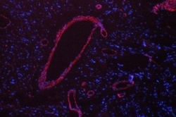
- Immunofluorescent staining of FFPE human glioma tissue with Calveolin-1 antibody (red) and DAPI nuclear counterstain (blue). HIER: boil tissue sections in pH8 EDTA buffer for 10-20 min followed by cooling at RT for 20 min.
Supportive validation
- Submitted by
- NSJ Bioreagents (provider)
- Main image
- Experimental details
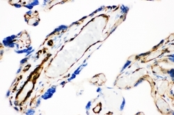
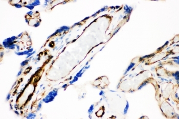
- IHC-P: Caveolin-1 antibody testing of human placenta tissue. HIER: steamed with pH6 citrate buffer.
- Submitted by
- NSJ Bioreagents (provider)
- Main image
- Experimental details
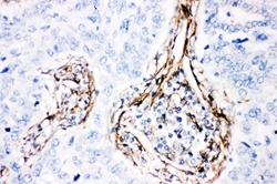
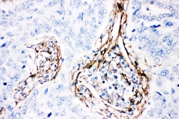
- IHC-P: Caveolin-1 antibody testing of human lung cancer tissue. HIER: steamed with pH6 citrate buffer.
- Submitted by
- NSJ Bioreagents (provider)
- Main image
- Experimental details
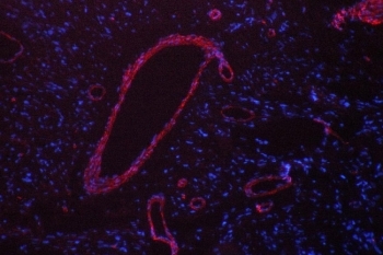
- Immunofluorescent staining of FFPE human glioma with Calveolin-1 antibody (red) and DAPI nuclear counterstain (blue). HIER: boil tissue sections in pH6, 10mM citrate buffer, for 10-20 min followed by cooling at RT for 20 min.

- Submitted by
- NSJ Bioreagents (provider)
- Main image
- Experimental details
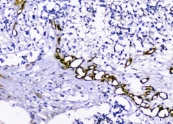
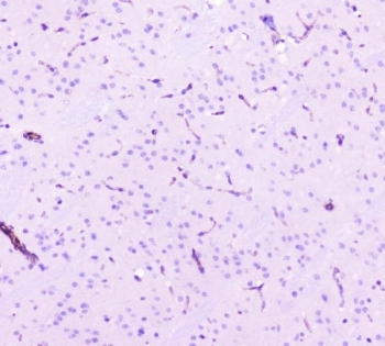
- IHC staining of FFPE human glioma tissue with Caveolin-1 antibody. HIER: boil tissue sections in pH8 EDTA for 20 min and allow to cool before testing.
- Submitted by
- NSJ Bioreagents (provider)
- Main image
- Experimental details
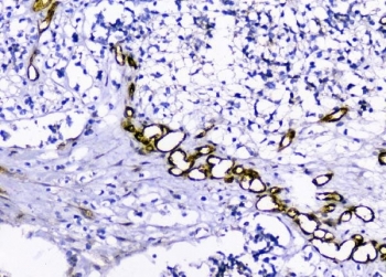
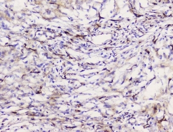
- IHC staining of FFPE human meningioma tissue with Caveolin-1 antibody. HIER: boil tissue sections in pH8 EDTA for 20 min and allow to cool before testing.
- Submitted by
- NSJ Bioreagents (provider)
- Main image
- Experimental details
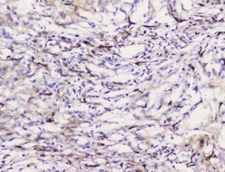
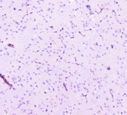
- IHC staining of FFPE mouse brain tissue with Caveolin-1 antibody. HIER: boil tissue sections in pH8 EDTA for 20 min and allow to cool before testing.
- Submitted by
- NSJ Bioreagents (provider)
- Main image
- Experimental details
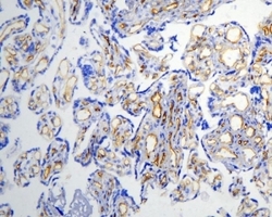
- IHC staining of frozen human placental tissue with Caveolin-1 antibody.

Supportive validation
- Submitted by
- NSJ Bioreagents (provider)
- Main image
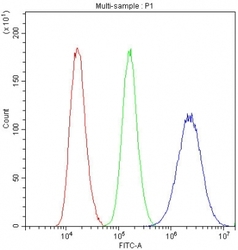
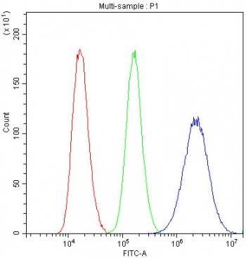
- Experimental details
- Flow cytometry testing of fixed and permeabilized human U-87 MG cells with Caveolin-1 antibody at 1ug/10^6 cells (blocked with goat sera); Red=cells alone, Green=isotype control, Blue= Caveolin-1 antibody.