 Explore
 Validate
 Learn
Learn Western blot
Antibody data
- Antibody Data
- Antigen structure
- References [2]
- Comments [0]
- Validations
- Western blot [4]
- Immunocytochemistry [2]
- Immunohistochemistry [1]
Submit
Validation data
Reference
Comment
Report error
- Product number
- PA1-37260 - Provider product page

- Provider
- Invitrogen Antibodies
- Product name
- Caveolin 1 Polyclonal Antibody
- Antibody type
- Polyclonal
- Antigen
- Synthetic peptide
- Description
- Heat-mediated antigen retrieval is recommended prior to staining, using a 10mM citrate buffer, pH 6.0, for 10 minutes followed by cooling at room temperature for 20 min. Following antigen retrieval, incubate samples with primary antibody for 10 min at room temperature. A suggested positive control is lung carcinoma.
- Reactivity
- Human
- Host
- Rabbit
- Isotype
- IgG
- Vial size
- 1 mL
- Storage
- Store at 4°C short term. For long term storage, store at -20°C, avoiding freeze/thaw cycles.
Submitted references Single-Cell RNA Sequencing of Childhood Ependymoma Reveals Neoplastic Cell Subpopulations That Impact Molecular Classification and Etiology.
Muscarinic receptor-mediated bronchoconstriction is coupled to caveolae in murine airways.
Gillen AE, Riemondy KA, Amani V, Griesinger AM, Gilani A, Venkataraman S, Madhavan K, Prince E, Sanford B, Hankinson TC, Handler MH, Vibhakar R, Jones KL, Mitra S, Hesselberth JR, Foreman NK, Donson AM
Cell reports 2020 Aug 11;32(6):108023
Cell reports 2020 Aug 11;32(6):108023
Muscarinic receptor-mediated bronchoconstriction is coupled to caveolae in murine airways.
Schlenz H, Kummer W, Jositsch G, Wess J, Krasteva G
American journal of physiology. Lung cellular and molecular physiology 2010 May;298(5):L626-36
American journal of physiology. Lung cellular and molecular physiology 2010 May;298(5):L626-36
No comments: Submit comment
Supportive validation
- Submitted by
- Invitrogen Antibodies (provider)
- Main image

- Experimental details
- Knockout of Caveolin 1 was achieved by CRISPR-Cas9 genome editing using LentiArray™ Lentiviral sgRNA (Product # A32042, Assay ID CRISPR639775_LV) and LentiArray Cas9 Lentivirus (Product # A32064). Western blot analysis of Caveolin 1 was performed by loading 30 µg of HeLa Cas9 (Lane 1), HeLa Cas9 treated with Palcitaxel (25 nM for 24 hrs) (Lane 2), HeLa Caveolin 1 KO (Lane 3) and HeLa Caveolin 1 KO treated with Palcitaxel (25 nM for 24 hrs) (Lane 4) whole cell extracts. The samples were electrophoresed using NuPAGE™ Novex™ 4-12% Bis-Tris Protein Gel (Product # NP0322BOX). Resolved proteins were then transferred onto a nitrocellulose membrane (Product # IB23001) by iBlot® 2 Dry Blotting System (Product # IB21001). The blot was probed with Anti-Caveolin 1 Polyclonal Antibody (Product # PA5-32297, 1:1,000 dilution) and Goat anti-Rabbit IgG (Heavy Chain) Superclonal™ Recombinant Secondary Antibody, HRP (Product # A27036, 1:4,000 dilution) using the iBright FL 1000 (Product # A32752). Chemiluminescent detection was performed using SuperSignal™ West Dura Extended Duration Substrate (Product # 34076). Loss of signal upon CRISPR mediated knockout (KO) using the LentiArray™ CRISPR product line confirms that antibody is specific to Caveolin 1.
- Submitted by
- Invitrogen Antibodies (provider)
- Main image
- Experimental details
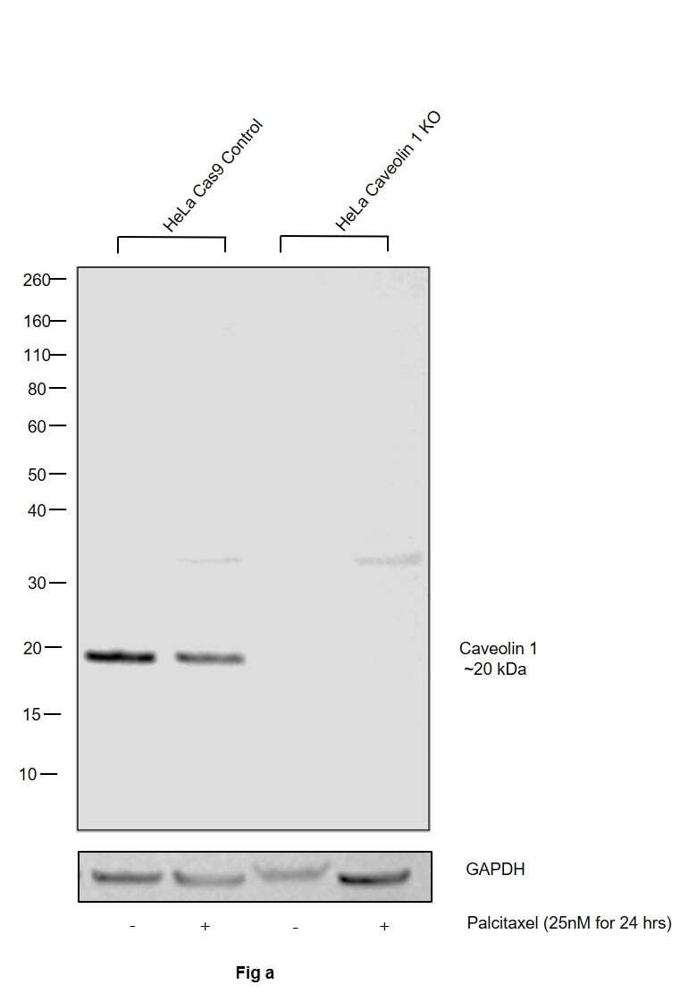
- Knockout of Caveolin 1 was achieved by CRISPR-Cas9 genome editing using LentiArray™ Lentiviral sgRNA (Product # A32042, Assay ID CRISPR639775_LV) and LentiArray Cas9 Lentivirus (Product # A32064). Western blot analysis of Caveolin 1 was performed by loading 30 µg of HeLa Cas9 (Lane 1), HeLa Cas9 treated with Palcitaxel (25 nM for 24 hrs) (Lane 2), HeLa Caveolin 1 KO (Lane 3) and HeLa Caveolin 1 KO treated with Palcitaxel (25 nMfor 24 hrs) (Lane 4) whole cell extracts. The samples were electrophoresed using NuPAGE™ Novex™ 4-12% Bis-Tris Protein Gel (Product # NP0322BOX). Resolved proteins were then transferred onto a nitrocellulose membrane (Product # IB23001) by iBlot® 2 Dry Blotting System (Product # IB21001). The blot was probed with Anti-Caveolin 1 Polyclonal Antibody (Product # PA1-37260, 1:1,000 dilution) and Goat anti-Rabbit IgG (Heavy Chain) Superclonal™ Recombinant Secondary Antibody, HRP (Product # A27036, 1:4,000 dilution) using the iBright FL 1000 (Product # A32752). Chemiluminescent detection was performed using SuperSignal™ West Dura Extended Duration Substrate (Product # 34076). Loss of signal upon CRISPR mediated knockout (KO) using the LentiArray™ CRISPR product line confirms that antibody is specific to Caveolin 1.

- Submitted by
- Invitrogen Antibodies (provider)
- Main image
- Experimental details
- Western blot was performed using Caveolin 1 Polyclonal Antibody (Product # PA5-32297) and a ~20 kDa band corresponding to Caveolin-1 was observed across the cell lines and tissues tested. Whole cell extracts (30 µg lysate) of A-431 (Lane 1), NIH/3T3 (Lane 2), IMR-32 (Lane 3), SH-SY5Y (Lane 4), Mouse Lung (Lane 5), Mouse Brown Fat (Lane 6), Rat Lung (Lane 7), Mouse Brain (Lane 8) were electrophoresed using Novex® NuPAGE® 4-12 % Bis-Tris gel (Product # NP0322BOX). Resolved proteins were then transferred onto a nitrocellulose membrane (Product # IB23001) by iBlot® 2 Dry Blotting System (Product # IB21001). The blot was probed with the primary antibody (1:1,000 dilution) and detected by chemiluminescence with Goat anti-Rabbit IgG (Heavy Chain) Superclonal™ Recombinant Secondary Antibody, HRP (Product # A27036, 1:4,000 dilution) using the iBright FL 1000 (Product # A32752). Chemiluminescent detection was performed using Novex® ECL Chemiluminescent Substrate Reagent Kit (Product # WP20005).
- Submitted by
- Invitrogen Antibodies (provider)
- Main image
- Experimental details
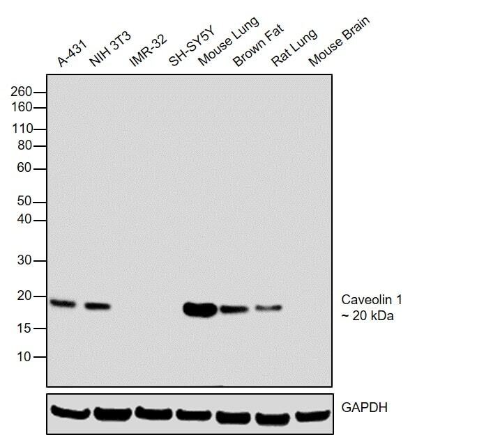
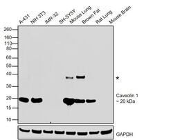
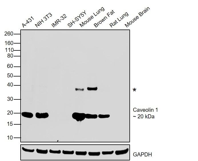
- Western blot was performed using Caveolin 1 Polyclonal Antibody (Product # PA1-37260) and an ~20 kDa band corresponding to Caveolin-1 was observed across the cell lines and tissues tested. An additional un-characterized band (*) was also observed. Whole cell extracts (30 µg lysate) of A-431 (Lane 1), NIH/3T3 (Lane 2), IMR-32 (Lane 3), SH-SY5Y (Lane 4), Mouse Lung (Lane 5), Mouse Brown Fat (Lane 6), Rat Lung (Lane 7), Mouse Brain (Lane 8) were electrophoresed using Novex® NuPAGE® 4-12 % Bis-Tris gel (Product # NP0322BOX). Resolved proteins were then transferred onto a nitrocellulose membrane (Product # IB23001) by iBlot® 2 Dry Blotting System (Product # IB21001). The blot was probed with the primary antibody (1:1,000 dilution) and detected by chemiluminescence with Goat anti-Rabbit IgG (Heavy Chain) Superclonal™ Recombinant Secondary Antibody, HRP (Product # A27036, 1:4,000 dilution) using the iBright FL 1000 (Product # A32752). Chemiluminescent detection was performed using Novex® ECL Chemiluminescent Substrate Reagent Kit (Product # WP20005).
Supportive validation
- Submitted by
- Invitrogen Antibodies (provider)
- Main image
- Experimental details
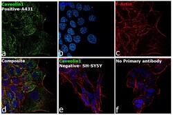
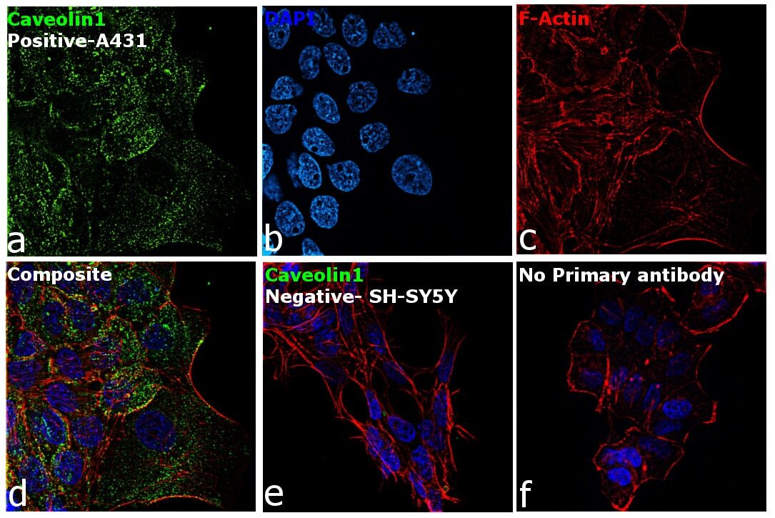
- Immunofluorescence analysis of Caveolin 1 was performed using 70% confluent log phase A-431 cells. The cells were fixed with 4% paraformaldehyde for 10 minutes, permeabilized with 0.1% Triton™ X-100 for 10 minutes, and blocked with 2% BSA for 1 hour at room temperature. The cells were labeled with Caveolin 1 Polyclonal Antibody (Product # PA5-32297) at 1:100 dilution in 0.1% BSA, incubated at 4 degree celsius overnight and then with Goat anti-Rabbit IgG (H+L) Highly Cross-Adsorbed Secondary Antibody, Alexa Fluor Plus 488 (Product # A32731) at a dilution of 1:2000 for 45 minutes at room temperature (Panel a: green). Nuclei (Panel b: blue) were stained with ProLong™ Diamond Antifade Mountant with DAPI (Product # P36962). F-actin (Panel c: red) was stained with Rhodamine Phalloidin (Product # R415, 1:300). Panel d represents the merged image showing nuclear localization. Panel e represents SH-SY5Y cells having no expression of Caveolin 1. Panel f represents control cells with no primary antibody to assess background. The images were captured at 60X magnification.

- Submitted by
- Invitrogen Antibodies (provider)
- Main image

- Experimental details
- Immunofluorescence analysis of Caveolin 1 was performed using 70% confluent log phase A-431 cells. The cells were fixed with 4% paraformaldehyde for 10 minutes, permeabilized with 0.1% Triton™ X-100 for 10 minutes, and blocked with 2% BSA for 1 hour at room temperature. The cells were labeled with Caveolin 1 Polyclonal Antibody (Product # PA5-32297) at 1:100 dilution in 0.1% BSA, incubated at 4 degree celsius overnight and then with Goat anti-Rabbit IgG (H+L) Highly Cross-Adsorbed Secondary Antibody, Alexa Fluor Plus 488 (Product # A32731) at a dilution of 1:2000 for 45 minutes at room temperature (Panel a: green). Nuclei (Panel b: blue) were stained with ProLong™ Diamond Antifade Mountant with DAPI (Product # P36962). F-actin (Panel c: red) was stained with Rhodamine Phalloidin (Product # R415, 1:300). Panel d represents the merged image showing nuclear localization. Panel e represents SH-SY5Y cells having no expression of Caveolin 1. Panel f represents control cells with no primary antibody to assess background. The images were captured at 60X magnification.
Supportive validation
- Submitted by
- Invitrogen Antibodies (provider)
- Main image
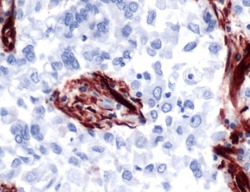
- Experimental details
- Immunohistochemical (paraffin) analysis of Caveolin-1 using anti-Caveolin-1 Polyclonal Antibody (Product # PA5-32297) in Lung Carcinoma Cancer Tissue. The recommended dilution for this antibody in immunohistochemistry applications is 1:100.
