Explore
 Validate
 Learn
LearnPA5-17447
antibody from Invitrogen Antibodies
Targeting: CAV1
CAV
 Western blot
 Immunocytochemistry
 Immunoprecipitation Immunohistochemistry Flow cytometry Other assay
Antibody data
- Antibody Data
- Antigen structure
- References [5]
- Comments [0]
- Validations
- Immunocytochemistry [8]
- Immunohistochemistry [2]
- Other assay [1]
Submit
Validation data
Reference
Comment
Report error
- Product number
- PA5-17447 - Provider product page

- Provider
- Invitrogen Antibodies
- Product name
- Caveolin 1 Polyclonal Antibody
- Antibody type
- Polyclonal
- Antigen
- Synthetic peptide
- Description
- It is not recommended to aliquot this antibody. This antibody is not cross-reactive with caveolin-2 or -3.
- Reactivity
- Human, Mouse, Rat, Bovine, Chicken/Avian, Hamster, Porcine, Zebrafish
- Host
- Rabbit
- Isotype
- IgG
- Vial size
- 100 μL
- Concentration
- 33 μg/mL
- Storage
- -20°C
Submitted references Immortalized human choroid plexus endothelial cells enable an advanced endothelial-epithelial two-cell type in vitro model of the choroid plexus.
Investigating receptor-mediated antibody transcytosis using blood-brain barrier organoid arrays.
Gut-Lung Dysbiosis Accompanied by Diabetes Mellitus Leads to Pulmonary Fibrotic Change through the NF-κB Signaling Pathway.
Caveolin-1 selectively regulates microRNA sorting into microvesicles after noxious stimuli.
Identification of a Pro-Angiogenic Potential and Cellular Uptake Mechanism of a LMW Highly Sulfated Fraction of Fucoidan from Ascophyllum nodosum.
Muranyi W, Schwerk C, Herold R, Stump-Guthier C, Lampe M, Fallier-Becker P, Weiß C, Sticht C, Ishikawa H, Schroten H
iScience 2022 Jun 17;25(6):104383
iScience 2022 Jun 17;25(6):104383
Investigating receptor-mediated antibody transcytosis using blood-brain barrier organoid arrays.
Simonneau C, Duschmalé M, Gavrilov A, Brandenberg N, Hoehnel S, Ceroni C, Lassalle E, Kassianidou E, Knoetgen H, Niewoehner J, Villaseñor R
Fluids and barriers of the CNS 2021 Sep 20;18(1):43
Fluids and barriers of the CNS 2021 Sep 20;18(1):43
Gut-Lung Dysbiosis Accompanied by Diabetes Mellitus Leads to Pulmonary Fibrotic Change through the NF-κB Signaling Pathway.
Wang G, Hu YX, He MY, Xie YH, Su W, Long D, Zhao R, Wang J, Dai C, Li H, Si ZP, Cheng X, Li RM, Li Z, Yang X
The American journal of pathology 2021 May;191(5):838-856
The American journal of pathology 2021 May;191(5):838-856
Caveolin-1 selectively regulates microRNA sorting into microvesicles after noxious stimuli.
Lee H, Li C, Zhang Y, Zhang D, Otterbein LE, Jin Y
The Journal of experimental medicine 2019 Sep 2;216(9):2202-2220
The Journal of experimental medicine 2019 Sep 2;216(9):2202-2220
Identification of a Pro-Angiogenic Potential and Cellular Uptake Mechanism of a LMW Highly Sulfated Fraction of Fucoidan from Ascophyllum nodosum.
Marinval N, Saboural P, Haddad O, Maire M, Bassand K, Geinguenaud F, Djaker N, Ben Akrout K, Lamy de la Chapelle M, Robert R, Oudar O, Guyot E, Laguillier-Morizot C, Sutton A, Chauvierre C, Chaubet F, Charnaux N, Hlawaty H
Marine drugs 2016 Oct 17;14(10)
Marine drugs 2016 Oct 17;14(10)
No comments: Submit comment
Supportive validation
- Submitted by
- Invitrogen Antibodies (provider)
- Main image

- Experimental details
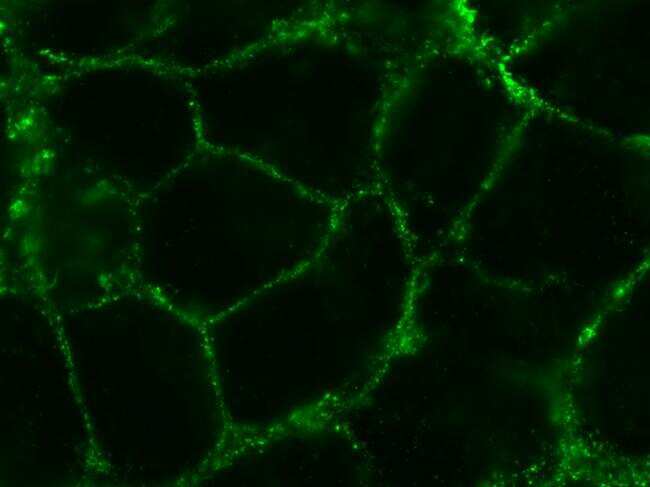
- Imunofluorescent analysis of caveolin-1 (green) in mouse kidney cells. Cells were fixed by methanol, incubated with a caveolin-1 rabbit polyclonal antibody (Product # PA5-17447) at 1:200 dilution, 4 degree overnight, then washed and incubated with goat anti rabbit conjugated with Alexa Fluor 488, at 1: 500 dilution, room temperature for 1 hour. The image was taken by zeiss Axioshop 40 microscope at 100 X condition. Note: Data courtesy of our Innovators Program.
- Submitted by
- Invitrogen Antibodies (provider)
- Main image
- Experimental details
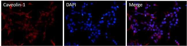
- Immunofluorescent analysis of Caveolin-1 (red) in HEK293T cells. Cells fixed with 4% formaldehyde were permeabilized and blocked with 1X PBS containing 5% BSA and 0.3% Triton X-100 for 1 hour at room temperature. Cells were probed with a Caveolin-1 polyclonal antibody (Product # PA5-17447) at a dilution of 1:100 overnight at 4°C in 1X PBS containing 1% BSA and 0.3% Triton X-100, washed with 1X PBS, and incubated with a fluorophore-conjugated goat anti-rabbit IgG secondary antibody at a dilution of 1:200 for 1 hour at room temperature. Nuclei (blue) were stained with DAPI. Images were taken on a Leica DM1000 microscope at 40X magnification. Data courtesy of the Innovators Program.
- Submitted by
- Invitrogen Antibodies (provider)
- Main image

- Experimental details
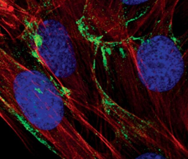
- Immunofluorescent analysis of Caveolin-1 in C2C12 cells using a Caveolin-1 polyclonal antibody (Product # PA5-17447) (green). Actin filaments are labeled with a fluorescent red phalloidin. DNA is labeled using a fluorescent blue dye.
- Submitted by
- Invitrogen Antibodies (provider)
- Main image

- Experimental details
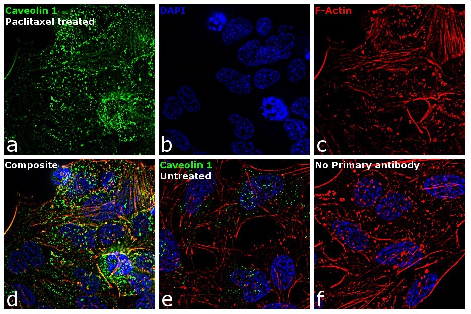
- Immunofluorescence analysis of Caveolin 1 was performed using 70% confluent log phase HeLa cells treated with 2 nM of Paclitaxel for 24 hr. The cells were fixed with 4% paraformaldehyde for 10 minutes, permeabilized with 0.1% Triton™ X-100 for 15 minutes, and blocked with 1% BSA for 1 hour at room temperature. The cells were labeled with Caveolin 1 Rabbit Polyclonal Antibody(Product # PA5-17447) at 1:100 dilution in 0.1% BSA, incubated at 4 degree Celsius overnight and then labeled with Goat anti-Rabbit IgG (H+L) Superclonal™ Secondary Antibody, Alexa Fluor® 488 conjugate (Product # A27034) at a dilution of 1:2000 for 45 minutes at room temperature (Panel a: green).Nuclei (Panel b: blue) were stained with ProLong™ Diamond Antifade Mountant with DAPI (Product # P36962). F-actin (Panel c: red) was stained with Rhodamine Phalloidin (Product # R415, 1:300). Panel d represents the merged image showing membrane localization. Panel e shows untreated cells with less membrane signal. Panel f represents control cells with no primary antibody to assess background. The images were captured at 60X magnification.
- Submitted by
- Invitrogen Antibodies (provider)
- Main image

- Experimental details
- Immunofluorescent analysis of Caveolin-1 in C2C12 cells using a Caveolin-1 polyclonal antibody (Product # PA5-17447) (green). Actin filaments are labeled with a fluorescent red phalloidin. DNA is labeled using a fluorescent blue dye.

- Submitted by
- Invitrogen Antibodies (provider)
- Main image

- Experimental details
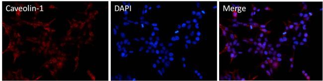
- Immunofluorescent analysis of Caveolin-1 (red) in HEK293T cells. Cells fixed with 4% formaldehyde were permeabilized and blocked with 1X PBS containing 5% BSA and 0.3% Triton X-100 for 1 hour at room temperature. Cells were probed with a Caveolin-1 polyclonal antibody (Product # PA5-17447) at a dilution of 1:100 overnight at 4°C in 1X PBS containing 1% BSA and 0.3% Triton X-100, washed with 1X PBS, and incubated with a fluorophore-conjugated goat anti-rabbit IgG secondary antibody at a dilution of 1:200 for 1 hour at room temperature. Nuclei (blue) were stained with DAPI. Images were taken on a Leica DM1000 microscope at 40X magnification. Data courtesy of the Innovators Program.
- Submitted by
- Invitrogen Antibodies (provider)
- Main image

- Experimental details
- Imunofluorescent analysis of caveolin-1 (green) in mouse kidney cells. Cells were fixed by methanol, incubated with a caveolin-1 rabbit polyclonal antibody (Product # PA5-17447) at 1:200 dilution, 4 degree overnight, then washed and incubated with goat anti rabbit conjugated with Alexa Fluor 488, at 1: 500 dilution, room temperature for 1 hour. The image was taken by zeiss Axioshop 40 microscope at 100 X condition. Note: Data courtesy of our Innovators Program.

- Submitted by
- Invitrogen Antibodies (provider)
- Main image

- Experimental details
- Immunofluorescence analysis of Caveolin 1 was performed using 70% confluent log phase HeLa cells treated with 2 nM of Paclitaxel for 24 hr. The cells were fixed with 4% paraformaldehyde for 10 minutes, permeabilized with 0.1% Triton™ X-100 for 15 minutes, and blocked with 1% BSA for 1 hour at room temperature. The cells were labeled with Caveolin 1 Rabbit Polyclonal Antibody(Product # PA5-17447) at 1:100 dilution in 0.1% BSA, incubated at 4 degree Celsius overnight and then labeled with Goat anti-Rabbit IgG (Heavy Chain) Superclonal™ Secondary Antibody, Alexa Fluor® 488 conjugate (Product # A27034) at a dilution of 1:2000 for 45 minutes at room temperature (Panel a: green).Nuclei (Panel b: blue) were stained with ProLong™ Diamond Antifade Mountant with DAPI (Product # P36962). F-actin (Panel c: red) was stained with Rhodamine Phalloidin (Product # R415, 1:300). Panel d represents the merged image showing membrane localization. Panel e shows untreated cells with less membrane signal. Panel f represents control cells with no primary antibody to assess background. The images were captured at 60X magnification.

Supportive validation
- Submitted by
- Invitrogen Antibodies (provider)
- Main image

- Experimental details
- Immunohistochemistry was performed on formaldehyde-fixed paraffin-embedded chicken embryo lung sections using an automated slide staining system. Heat-induced epitope retrieval was performed for 60 minutes at 100°C to expose target antigens. Tissues were stained with a Caveolin-1 polyclonal antibody (Product # PA5-17447) at a dilution of 1:100 for at least 30 minutes. Tissues were counterstained with Hematoxylin and visualized by light microscopy. Magnification = 40X. Data courtesy of the Innovators Program.

- Submitted by
- Invitrogen Antibodies (provider)
- Main image

- Experimental details
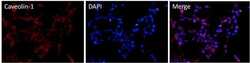
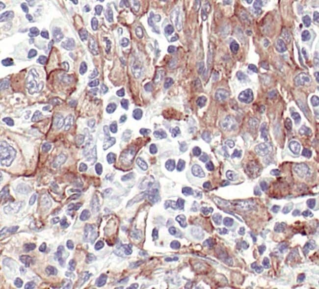
- Immunohistochemistry was performed on paraffin-embedded human lung carcinoma. To expose target proteins, antigen retrieval method was performed by heating tissues in 10mM sodium citrate buffer pH 6.0 for 10 minutes. Following antigen retrieval, tissues were treated with 3% hydrogen peroxide to quench endogenous peroxidase activity, blocked with 5% normal goat serum in TBST for 1 hour at room temperature, and then probed with a Caveolin-1 polyclonal antibody (Product # PA5-17447) at a dilution of 1:250 overnight at 4°C in a humidified chamber. Detection was performed using an HRP-conjugated detection reagent followed by DAB substrate.
Supportive validation
- Submitted by
- Invitrogen Antibodies (provider)
- Main image

- Experimental details
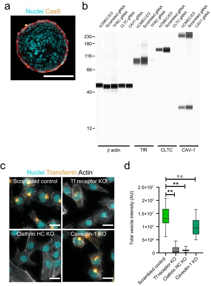
- Fig. 4 Establishment and characterization of Cas9 brain endothelial cell lines. a Representative confocal image acquired at the core of a blood-barrier organoid assembled with brain endothelial cells expressing Cas9 (orange). Cas9 brain endothelial cells localize only at the periphery of blood-brain barrier organoids. Nuclei labelled with DAPI (cyan). Scale bar, 100 mum. b Representative Western blot image showing the expression of Transferrin receptor, clathrin heavy chain or caveolin-1 in hCMEC/D3 brain endothelial cells expressing Cas9 and transduced with either scrambled gRNA or gRNA against the target gene. beta actin expression is shown as a reference control gene. c Representative fluorescent images of hCMEC/D3 brain endothelial Cas9 or knockout cells after incubation with fluorescently labelled transferrin (yellow) for 30 min. Actin is labelled with phalloidin (grey) to visualize cell contours and nuclei are labelled with DAPI (cyan). Scale bars, 20 mum. d Quantification of transferrin internalization in hCMEC/D3 Cas9 or knockout cells. Graph shows boxplots with interquartile ranges and median. Lines show the 5th and 95th percentiles. Differences between the scrambled control and transferrin receptor or clathrin heavy-chain knockout cells were statistically significant (**p = 0.018) whereas the difference between the scrambled control and caveolin-1 knockout cells was not statistically significant (p = 0.418). Comparisons were evaluated by one-way ANOVA followed by Du