Explore
 Validate
 Learn
Learn Western blot
Antibody data
- Antibody Data
- Antigen structure
- References [1]
- Comments [0]
- Validations
- Western blot [3]
- Immunohistochemistry [1]
Submit
Validation data
Reference
Comment
Report error
- Product number
- AF5736 - Provider product page

- Provider
- R&D Systems
- Product name
- Human Caveolin-1 Antibody
- Antibody type
- Polyclonal
- Description
- Antigen Affinity-purified. Detects human Caveolin-1 in Western blots.
- Reactivity
- Human
- Host
- Goat
- Conjugate
- Unconjugated
- Antigen sequence
Q03135- Isotype
- IgG
- Vial size
- 100 ug
- Concentration
- LYOPH
- Storage
- Use a manual defrost freezer and avoid repeated freeze-thaw cycles. 12 months from date of receipt, -20 to -70 °C as supplied. 1 month, 2 to 8 °C under sterile conditions after reconstitution. 6 months, -20 to -70 °C under sterile conditions after reconstitution.
Submitted references Snail-Induced Epithelial-to-Mesenchymal Transition Enhances P-gp-Mediated Multidrug Resistance in HCC827 Cells.
Tomono T, Yano K, Ogihara T
Journal of pharmaceutical sciences 2017 Sep;106(9):2642-2649
Journal of pharmaceutical sciences 2017 Sep;106(9):2642-2649
No comments: Submit comment
Supportive validation
- Submitted by
- R&D Systems (provider)
- Main image

- Experimental details
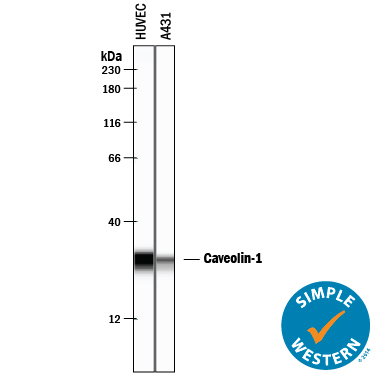
- Detection of Human Caveolin-1 by Simple WesternTM. Simple Western lane view shows lysates of HUVEC human umbilical vein endothelial cells and A431 human epithelial carcinoma cell line, loaded at 0.2 mg/mL. A specific band was detected for Caveolin-1 at approximately 29 kDa (as indicated) using 2 µg/mL of Goat Anti-Human Caveolin-1 Antigen Affinity-purified Polyclonal Antibody (Catalog # AF5736) followed by 1:50 dilution of HRP-conjugated Anti-Goat IgG Secondary Antibody (Catalog # HAF109). This experiment was conducted under reducing conditions and using the 12-230 kDa separation system.
- Submitted by
- R&D Systems (provider)
- Main image
- Experimental details
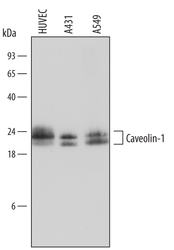
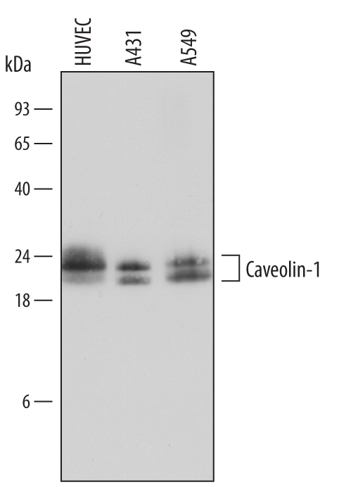
- Detection of Human Caveolin-1 by Western Blot. Western blot shows lysates of HUVEC human umbilical vein endothelial cells, A431 human epithelial carcinoma cell line, and A549 human lung carcinoma cell line. PVDF Membrane was probed with 0.2 µg/mL of Goat Anti-Human Caveolin-1 Antigen Affinity-purified Polyclonal Antibody (Catalog # AF5736) followed by HRP-conjugated Anti-Goat IgG Secondary Antibody (Catalog # HAF109). Specific bands were detected for Caveolin-1 at approximately 21 to 24 kDa (as indicated). This experiment was conducted under reducing conditions and using Immunoblot Buffer Group 1.
- Submitted by
- R&D Systems (provider)
- Main image

- Experimental details
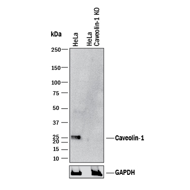
- Western Blot Shows Human Caveolin-1 Specificity by Using Knockout Cell Line. Western blot shows lysates of HeLa human cervical epithelial carcinoma parental cell line and Caveolin-1 knockout HeLa cell line (KO). PVDF membrane was probed with 0.2 µg/mL of Goat Anti-Human Caveolin-1 Antigen Affinity-purified Polyclonal Antibody (Catalog # AF5736) followed by HRP-conjugated Anti-Goat IgG Secondary Antibody (Catalog # HAF017). A specific band was detected for Caveolin-1 at approximately 25 kDa (as indicated) in the parental HeLa cell line, but is not detectable in knockout HeLa cell line. GAPDH (Catalog # AF5718) is shown as a loading control. This experiment was conducted under reducing conditions and using Immunoblot Buffer Group 1.
Supportive validation
- Submitted by
- R&D Systems (provider)
- Main image

- Experimental details
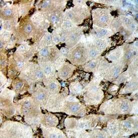
- Caveolin-1 in Human Liver. Caveolin-1 was detected in immersion fixed paraffin-embedded sections of human liver using Goat Anti-Human Caveolin-1 Antigen Affinity-purified Polyclonal Antibody (Catalog # AF5736) at 10 µg/mL overnight at 4 °C. Before incubation with the primary antibody, tissue was subjected to heat-induced epitope retrieval using Antigen Retrieval Reagent-Basic (Catalog # CTS013). Tissue was stained using the Anti-Goat HRP-DAB Cell & Tissue Staining Kit (brown; Catalog # CTS008) and counterstained with hematoxylin (blue). Specific staining was localized to endothelial cells in bile canaliculi. View our protocol for Chromogenic IHC Staining of Paraffin-embedded Tissue Sections.