Explore
 Validate
 Learn
Learn Western blot
Antibody data
- Antibody Data
- Antigen structure
- References [1]
- Comments [0]
- Validations
- Western blot [3]
- Immunohistochemistry [1]
Submit
Validation data
Reference
Comment
Report error
- Product number
- MAB4299 - Provider product page

- Provider
- R&D Systems
- Product name
- Human IkB-alpha Antibody
- Antibody type
- Monoclonal
- Description
- Protein A or G purified from hybridoma culture supernatant. Detects human IkB-alpha in Western blots.
- Reactivity
- Human
- Host
- Mouse
- Conjugate
- Unconjugated
- Antigen sequence
P25963- Isotype
- IgG
- Antibody clone number
- 417208
- Vial size
- 50 ug
- Concentration
- LYOPH
- Storage
- Use a manual defrost freezer and avoid repeated freeze-thaw cycles. 12 months from date of receipt, -20 to -70 °C as supplied. 1 month, 2 to 8 °C under sterile conditions after reconstitution. 6 months, -20 to -70 °C under sterile conditions after reconstitution.
Submitted references ERK5 protein promotes, whereas MEK1 protein differentially regulates, the Toll-like receptor 2 protein-dependent activation of human endothelial cells and monocytes.
Wilhelmsen K, Mesa KR, Lucero J, Xu F, Hellman J
The Journal of biological chemistry 2012 Aug 3;287(32):26478-94
The Journal of biological chemistry 2012 Aug 3;287(32):26478-94
No comments: Submit comment
Supportive validation
- Submitted by
- R&D Systems (provider)
- Main image
- Experimental details
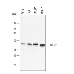
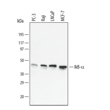
- Detection of Human IkB-alpha by Western Blot. Western blot shows lysates of Raji human Burkitt's lymphoma cell line, MCF-7 human breast cancer cell line, PC-3 human prostate cancer cell line, and LNCaP human prostate cancer cell line. PVDF membrane was probed with 0.1 µg/mL of Mouse Anti-Human IkB-alpha Monoclonal Antibody (Catalog # MAB4299) followed by HRP-conjugated Anti-Mouse IgG Secondary Antibody (Catalog # HAF007). A specific band was detected for IkB-alpha at approximately 44 kDa (as indicated). This experiment was conducted under reducing conditions and using Immunoblot Buffer Group 4.
- Submitted by
- R&D Systems (provider)
- Main image
- Experimental details
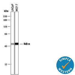
- Detection of Human IkB-alpha by Simple WesternTM. Simple Western lane view shows lysates of LNCaP human prostate cancer cell line and MCF-7 human breast cancer cell line, loaded at 0.5 mg/mL. A specific band was detected for IkB-alpha at approximately 44 kDa (as indicated) using 1 µg/mL of Mouse Anti-Human IkB-alpha Monoclonal Antibody (Catalog # MAB4299). This experiment was conducted under reducing conditions and using the 12-230 kDa separation system.

- Submitted by
- R&D Systems (provider)
- Main image
- Experimental details
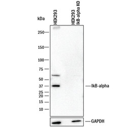
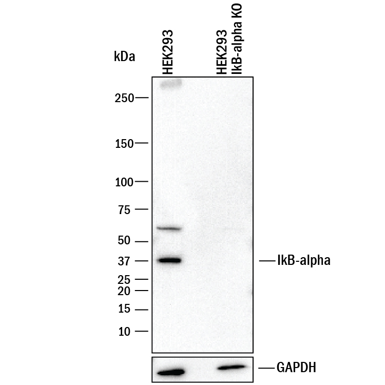
- Western Blot Shows Human IkB-alpha Specificity by Using Knockout Cell Line. Western blot shows lysates of HEK293T human embryonic kidney parental cell line and IkB-alpha knockout HEK293T cell line (KO). PVDF membrane was probed with 0.1 µg/mL of Mouse Anti-Human IkB-alpha Monoclonal Antibody (Catalog # MAB4299) followed by HRP-conjugated Anti-Mouse IgG Secondary Antibody (Catalog # HAF018). A specific band was detected for IkB-alpha at approximately 38 kDa (as indicated) in the parental HEK293T cell line, but is not detectable in knockout HEK293T cell line. GAPDH (Catalog # MAB5718) is shown as a loading control. This experiment was conducted under reducing conditions and using Immunoblot Buffer Group 1.
Supportive validation
- Submitted by
- R&D Systems (provider)
- Main image

- Experimental details
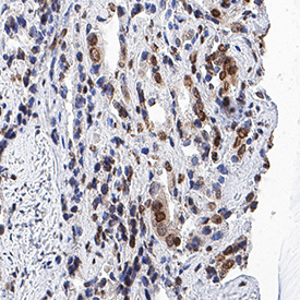
- IkB-alpha in Human Prostate Cancer Tissue. IkB-alpha was detected in immersion fixed paraffin-embedded sections of human prostate cancer tissue using Mouse Anti-Human IkB-alpha Monoclonal Antibody (Catalog # MAB4299) at 5 µg/mL for 1 hour at room temperature followed by incubation with the Anti-Mouse IgG VisUCyte™ HRP Polymer Antibody (Catalog # VC001). Tissue was stained using DAB (brown) and counterstained with hematoxylin (blue). Specific staining was localized to cytoplasm and nuclei in cancer cells. View our protocol for IHC Staining with VisUCyte HRP Polymer Detection Reagents.