Explore
 Validate
 Learn
Learn Western blot
 ELISA
ELISAAntibody data
- Antibody Data
- Antigen structure
- References [0]
- Comments [0]
- Validations
- Western blot [1]
- Immunocytochemistry [2]
Submit
Validation data
Reference
Comment
Report error
- Product number
- LS-C153771 - Provider product page

- Provider
- LSBio
- Product name
- RELA / NFKB p65 Antibody (C-Terminus, clone 27F9.G4) LS-C153771
- Antibody type
- Monoclonal
- Description
- Protein A affinity chromatography
- Reactivity
- Human
- Host
- Mouse
- Isotype
- IgG
- Antibody clone number
- 27F9.G4
- Storage
- Short term: store at 4°C. Long term: aliquot and store at -20°C. Avoid freeze-thaw cycles. Store undiluted.
No comments: Submit comment
Supportive validation
- Submitted by
- LSBio (provider)
- Enhanced method
- Genetic validation
- Main image

- Experimental details
- anti NFKB p65 (Rel A) monoclonal antibody Anti-NFKB p65 (Rel A) (MOUSE) Monoclonal Antibody - 200-301-065 was used to detect ~65 kD band (red arrow) in HeLa whole cell lysate. Lysate was run on 4-20% gradient gel transferred under standard conditions and blocked in 1% BSA-TTBS 30 min RT. Blot was probed with monoclonal anti p65 1:1000 in 1% BSA-TBS-T o/n 4? and detected with HRP conjugated Rb-anti Mouse antibody 1:40000 in MB-070 30 min RT.

Supportive validation
- Submitted by
- LSBio (provider)
- Enhanced method
- Genetic validation
- Main image

- Experimental details
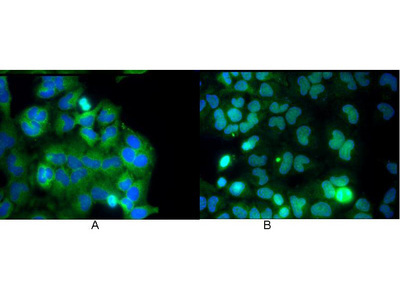
- Monoclonal anti NFKB p65 (Rel A) antibody was used to detect p65 by immunofluorescence at a dilution of 1:5000. HeLa cells were grown to sub-confluent on 18 mm2 glass coverslips #1.5. Cells were either unstimulated (A), or stimulated (B) with 50 ng/ml of TNF alpha for 30 min prior fixation. Cells were then fixed in methanol and blocked with 10% normal goat serum (NGS), in PBS, and Triton X 0.2% (Tx) and incubated for 1 hr at RT with primary ab, counterstained with DAPI and washed in PBS/NGS/Tx. Cells were incubated for 1 hr at RT with Atto 425 conjugated anti mouse secondary antibody for STED CW imaging. Data was collected on a STED-CW TCS-SP5 Confocal system equipped with a DFC 350FX camera allowing sequential acquisition in wide field, confocal and STED CW imaging on the same system.

- Submitted by
- LSBio (provider)
- Main image

- Experimental details
- Monoclonal anti NFKB p65 (Rel A) antibody was used to detect p65 by immunofluorescence at a dilution of 1:5000. HeLa cells were grown to sub-confluent on 18 mm2 glass coverslips #1.5. Cells were either unstimulated (A), or stimulated (B) with 50 ng/ml of TNF alpha for 30 min prior fixation. Cells were then fixed in methanol and blocked with 10% normal goat serum (NGS), in PBS, and Triton X 0.2% (Tx) and incubated for 1 hr at RT with primary ab, counterstained with DAPI and washed in PBS/NGS/Tx. Cells were incubated for 1 hr at RT with Atto 425 conjugated anti mouse secondary antibody for STED CW imaging. Data was collected on a STED-CW TCS-SP5 Confocal system equipped with a DFC 350FX camera allowing sequential acquisition in wide field, confocal and STED CW imaging on the same system.