Explore
 Validate
 Learn
Learn Western blot
 ELISA
ELISA Immunohistochemistry
Antibody data
- Antibody Data
- Antigen structure
- References [0]
- Comments [0]
- Validations
- Western blot [4]
Submit
Validation data
Reference
Comment
Report error
- Product number
- LS-B652 - Provider product page

- Provider
- LSBio
- Product name
- IHC-plus™ RELA / NFKB p65 Antibody (phospho-Ser529) LS-B652
- Antibody type
- Polyclonal
- Description
- Delipidated and defibrinated
- Reactivity
- Human
- Host
- Rabbit
- Storage
- Store at 4°C or -20°C. Avoid freeze-thaw cycles.
No comments: Submit comment
Supportive validation
- Submitted by
- LSBio (provider)
- Enhanced method
- Genetic validation
- Main image
- Experimental details
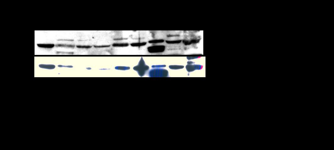
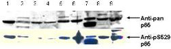
- Top row is anti-pan p65. Bottom row is anti-pS529 showing phospho p65 staining in carcinoma cells. Immunoblot of total protein lysates from various human head and neck tumors shows phospho p65 staining in tumor cell lines using phospho specific polyclonal anti-human pS529 p65. Lanes 1-6 contain protein lysates from human squamal carcinoma cell lines. Lane 7 is a protein lysate from a primary culture of human keratinocytes. Lane 8 contains protein lysate from ATCC SCC9 cells (also a head and
- Submitted by
- LSBio (provider)
- Enhanced method
- Genetic validation
- Main image
- Experimental details
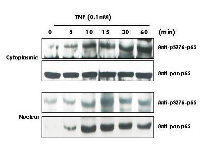
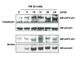
- Anti-Human pS276 p65 Antibody - Western Blot. TNF Induces phosphorylation of p65 in KBM-5 cells.

- Submitted by
- LSBio (provider)
- Enhanced method
- Genetic validation
- Main image
- Experimental details
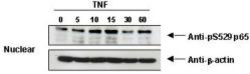
- Anti-pS529 shows phospho p65 staining in carcinoma cells.

- Submitted by
- LSBio (provider)
- Enhanced method
- Genetic validation
- Main image
- Experimental details
- Anti-Human pS276 p65 Antibody - Western Blot. TNF Induces phosphorylation of p65 in KBM-5 cells. Cytoplasmic and nuclear protein lysates prepared after 0, 5, 10, 15, 30 and 60 minutes of 0.1 nM TNF treatment of KBM-5 cells shows inducible phosphorylation using phospho specific polyclonal anti-human pS276 p65. pan reactive anti-p65 (code# LS-B653) was used a control to show the presence of total p65 in both the cytoplasmic and nuclear extracts. Phosphorylation of p65 occurs after approximately 10 min of TNF exposure. Migration of phosphorylated p65 into the nucleus occurs within a similar time frame. HRP conjugated Gt-anti-Rabbit IgG was used to develop the Western blot of a chemiluminescent detection method. Other detection methods will yield similar results. Personal Communication, Aggarwal BB.