 Explore
 Validate
 Learn
Learn Western blot
Antibody data
- Antibody Data
- Antigen structure
- References [2]
- Comments [0]
- Validations
- Western blot [2]
- Immunocytochemistry [1]
Submit
Validation data
Reference
Comment
Report error
- Product number
- MAB72261 - Provider product page

- Provider
- R&D Systems
- Product name
- Human Phospho-RelA/NFkB p65 (S536) Antibody
- Antibody type
- Monoclonal
- Description
- Protein A or G purified from cell culture supernatant. Detects human RelA/NF kappa B p65 when phosphorylated at S536.
- Reactivity
- Human
- Host
- Rabbit
- Conjugate
- Unconjugated
- Isotype
- IgG
- Antibody clone number
- 1091B
- Vial size
- 100 ug
- Storage
- Use a manual defrost freezer and avoid repeated freeze-thaw cycles. 12 months from date of receipt, -20 to -70 °C as supplied. 1 month, 2 to 8 °C under sterile conditions after reconstitution. 6 months, -20 to -70 °C under sterile conditions after reconstitution.
Submitted references Tumor associated macrophages induce epithelial to mesenchymal transition via the EGFR/ERK1/2 pathway in head and neck squamous cell carcinoma.
HIV-1 Tat protein induces DNA damage in human peripheral blood B-lymphocytes via mitochondrial ROS production.
Gao L, Zhang W, Zhong WQ, Liu ZJ, Li HM, Yu ZL, Zhao YF
Oncology reports 2018 Nov;40(5):2558-2572
Oncology reports 2018 Nov;40(5):2558-2572
HIV-1 Tat protein induces DNA damage in human peripheral blood B-lymphocytes via mitochondrial ROS production.
El-Amine R, Germini D, Zakharova VV, Tsfasman T, Sheval EV, Louzada RAN, Dupuy C, Bilhou-Nabera C, Hamade A, Najjar F, Oksenhendler E, Lipinski M, Chernyak BV, Vassetzky YS
Redox biology 2018 May;15:97-108
Redox biology 2018 May;15:97-108
No comments: Submit comment
Supportive validation
- Submitted by
- R&D Systems (provider)
- Main image
- Experimental details
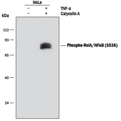
- Detection of Human Phospho-RelA/NF kappa B p65 (S536) by Western Blot. Western blot shows lysates of HeLa human cervical epithelial carcinoma cell line untreated (-) or treated (+) with 100 nM Calyculin A (Catalog # 1336) and 20 ng/mL Recombinant Human TNF-alpha (Catalog # 210-TA) for 10 minutes. PVDF membrane was probed with 0.1 μg/mL of Rabbit Anti-Human Phospho-RelA/NF kappa B p65 (S536) Monoclonal Antibody (Catalog # MAB72261) followed by HRP-conjugated Anti-Rabbit IgG Secondary Antibody (Catalog # HAF008). A specific band was detected for Phospho-RelA/NF kappa B p65 (S536) at approximately 65 kDa (as indicated). This experiment was conducted under reducing conditions and using Immunoblot Buffer Group 1.

- Submitted by
- R&D Systems (provider)
- Main image

- Experimental details
- Detection of Human Phospho-RelA/NF kappa B p65 (S536) by Simple WesternTM. Simple Western lane view shows lysates of HeLa human cervical epithelial carcinoma cell line untreated (-) or treated (+) with 100 nM Calyculin A (Catalog # 1336) and 20 ng/mL Recombinant Human TNF-alpha (Catalog # 210-TA) for 10 minutes, loaded at 0.2 mg/mL. A specific band was detected for Phospho-RelA/NF kappa B p65 (S536) at approximately 66 kDa (as indicated) using 1.0 µg/mL of Rabbit Anti-Human Phospho-RelA/NF kappa B p65 (S536) Monoclonal Antibody (Catalog # MAB72261). This experiment was conducted under reducing conditions and using the 12-230 kDa separation system. Non-specific interaction with the 230 kDa Simple Western standard may be seen with this antibody.

Supportive validation
- Submitted by
- R&D Systems (provider)
- Main image
- Experimental details
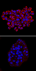
- Phospho-RelA/NFkB p65 (S536) in HT-29 Human Cell Line. RelA/NF kappa B p65 phosphorylated at S536 was detected in immersion fixed HT-29 human colon adenocarcinoma cell line untreated (lower panel) or treated with Calyculin A (Catalog # 1336) and Recombinant Human TNF-alpha (Catalog # 210-TA; upper panel) using Rabbit Anti-Human Phospho-RelA/NF kappa B p65 (S536) Monoclonal Antibody (Catalog # MAB72261) at 10 μg/mL for 3 hours at room temperature. Cells were stained using the NorthernLights™ 557-conjugated Anti-Rabbit IgG Secondary Antibody (red; Catalog # NL004) and counterstained with DAPI (blue). Specific staining was localized to cytoplasm and cell surfaces. View our protocol for Fluorescent ICC Staining of Cells on Coverslips.
